[English] 日本語
Yorodumi
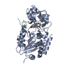
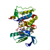
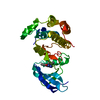
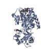
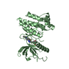
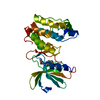
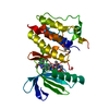
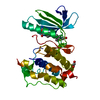
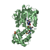
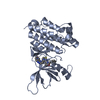
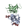
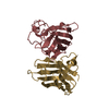
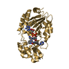
- PDB-3wzj: CRYSTAL STRUCTURE OF HUMAN MPS1 CATALYTIC DOMAIN IN COMPLEX WITH ... -

+
Open data


ID or keywords:

Loading...

-
Basic information

Entry
Database: PDB / ID: 3wzj
TitleCRYSTAL STRUCTURE OF HUMAN MPS1 CATALYTIC DOMAIN IN COMPLEX WITH 4-(6-(cyclohexylamino)-8-(((tetrahydro-2H-pyran-4-yl)methyl)amino)imidazo[1,2-b]pyridazin-3-yl)-N-cyclopropylbenzamide
ComponentsDual specificity protein kinase TTK
KeywordsTRANSFERASE / ATP Binding / Phosphorylation
Function / homology
Function and homology information


protein localization to meiotic spindle midzone / meiotic spindle assembly checkpoint signaling / kinetochore binding / female meiosis chromosome segregation / protein localization to kinetochore / dual-specificity kinase / spindle organization / mitotic spindle assembly checkpoint signaling / protein serine/threonine/tyrosine kinase activity / mitotic spindle organization ...protein localization to meiotic spindle midzone / meiotic spindle assembly checkpoint signaling / kinetochore binding / female meiosis chromosome segregation / protein localization to kinetochore / dual-specificity kinase / spindle organization / mitotic spindle assembly checkpoint signaling / protein serine/threonine/tyrosine kinase activity / mitotic spindle organization / chromosome segregation / spindle / kinetochore / protein tyrosine kinase activity / phosphorylation / protein serine kinase activity / protein serine/threonine kinase activity / positive regulation of cell population proliferation / ATP binding / membrane / identical protein binding / nucleus / cytoplasm
Similarity search - Function
Protein kinase Mps1 family / Tetratricopeptide-like helical domain superfamily / Transferase(Phosphotransferase) domain 1 / Transferase(Phosphotransferase); domain 1 / Phosphorylase Kinase; domain 1 / Phosphorylase Kinase; domain 1 / Serine/threonine-protein kinase, active site / Serine/Threonine protein kinases active-site signature. / Protein kinase domain / Serine/Threonine protein kinases, catalytic domain ...Protein kinase Mps1 family / Tetratricopeptide-like helical domain superfamily / Transferase(Phosphotransferase) domain 1 / Transferase(Phosphotransferase); domain 1 / Phosphorylase Kinase; domain 1 / Phosphorylase Kinase; domain 1 / Serine/threonine-protein kinase, active site / Serine/Threonine protein kinases active-site signature. / Protein kinase domain / Serine/Threonine protein kinases, catalytic domain / Protein kinase, ATP binding site / Protein kinases ATP-binding region signature. / Protein kinase domain profile. / Protein kinase domain / Protein kinase-like domain superfamily / 2-Layer Sandwich / Orthogonal Bundle / Mainly Alpha / Alpha Beta
Similarity search - Domain/homology
Chem-O43 / Dual specificity protein kinase TTK
Similarity search - Component
Biological speciesHomo sapiens (human)
MethodX-RAY DIFFRACTION / MOLECULAR REPLACEMENT / molecular replacement / Resolution: 2.75 Å
AuthorsKusakabe, K. / Ide, N. / Daigo, Y. / Itoh, T. / Yamamoto, T. / Kojima, E. / Mitsuoka, Y. / Tadano, G. / Tagashira, S. / Higashino, K. ...Kusakabe, K. / Ide, N. / Daigo, Y. / Itoh, T. / Yamamoto, T. / Kojima, E. / Mitsuoka, Y. / Tadano, G. / Tagashira, S. / Higashino, K. / Okano, Y. / Sato, Y. / Inoue, M. / Iguchi, M. / Kanazawa, T. / Ishioka, Y. / Dohi, K. / Kido, Y. / Sakamoto, S. / Ando, S. / Maeda, M. / Higaki, M. / Yoshizawa, H. / Mura, H. / Nakamura, Y.
CitationJournal: J.Med.Chem. / Year: 2015
Title: Discovery of imidazo[1,2-b]pyridazine derivatives: selective and orally available Mps1 (TTK) kinase inhibitors exhibiting remarkable antiproliferative activity.
Authors: Kusakabe, K. / Ide, N. / Daigo, Y. / Itoh, T. / Yamamoto, T. / Hashizume, H. / Nozu, K. / Yoshida, H. / Tadano, G. / Tagashira, S. / Higashino, K. / Okano, Y. / Sato, Y. / Inoue, M. / ...Authors: Kusakabe, K. / Ide, N. / Daigo, Y. / Itoh, T. / Yamamoto, T. / Hashizume, H. / Nozu, K. / Yoshida, H. / Tadano, G. / Tagashira, S. / Higashino, K. / Okano, Y. / Sato, Y. / Inoue, M. / Iguchi, M. / Kanazawa, T. / Ishioka, Y. / Dohi, K. / Kido, Y. / Sakamoto, S. / Ando, S. / Maeda, M. / Higaki, M. / Baba, Y. / Nakamura, Y.
History
DepositionSep 29, 2014Deposition site: PDBJ / Processing site: PDBJ
Revision 1.0Feb 11, 2015Provider: repository / Type: Initial release
Revision 1.1Nov 22, 2017Group: Refinement description / Category: software
Revision 1.2Aug 24, 2022Group: Database references / Derived calculations
Category: citation / citation_author ...citation / citation_author / database_2 / struct_ref_seq_dif / struct_site
Item: _citation.journal_volume / _citation.page_first ..._citation.journal_volume / _citation.page_first / _citation.page_last / _citation.title / _citation_author.name / _database_2.pdbx_DOI / _database_2.pdbx_database_accession / _struct_ref_seq_dif.details / _struct_site.pdbx_auth_asym_id / _struct_site.pdbx_auth_comp_id / _struct_site.pdbx_auth_seq_id

-
Structure visualization

Structure viewerMolecule:
MolmilJmol/JSmol

Downloads & links

-
Assembly

Deposited unit
A: Dual specificity protein kinase TTK
hetero molecules


Theoretical massNumber of molelcules
Total (without water)37,3472
Polymers36,8581
Non-polymers4891
Water362
1


  • Idetical with deposited unit
  • defined by author
TypeNameSymmetry operationNumber
identity operation1_555x,y,z1
2
A: Dual specificity protein kinase TTK
hetero molecules

A: Dual specificity protein kinase TTK
hetero molecules


Theoretical massNumber of molelcules
Total (without water)74,6944
Polymers73,7172
Non-polymers9772
Water362
TypeNameSymmetry operationNumber
identity operation1_555x,y,z1
crystal symmetry operation2_455-x-1,-y,z1
Buried area1280 Å2
ΔGint-13 kcal/mol
Surface area23700 Å2
MethodPISA
Unit cell
Length a, b, c (Å)70.961, 110.389, 114.356
Angle α, β, γ (deg.)90.000, 90.000, 90.000
Int Tables number23
Space group name H-MI222

-
Components

#1: Protein Dual specificity protein kinase TTK / Phosphotyrosine picked threonine-protein kinase / PYT


Mass: 36858.363 Da / Num. of mol.: 1 / Fragment: MPS1 KINASE DOMAIN, UNP residues 516-820
Source method: isolated from a genetically manipulated source
Source: (gene. exp.) Homo sapiens (human) / Gene: TTK, MPS1, MPS1L1 / Production host: CELL-FREE SYNTHESIS (others) / References: UniProt: P33981, dual-specificity kinase
#2: Chemical ChemComp-O43 / 4-{6-(cyclohexylamino)-8-[(tetrahydro-2H-pyran-4-ylmethyl)amino]imidazo[1,2-b]pyridazin-3-yl}-N-cyclopropylbenzamide


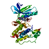
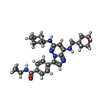
Mass: 488.624 Da / Num. of mol.: 1 / Source method: obtained synthetically / Formula: C28H36N6O2
#3: Water ChemComp-HOH / water / Water


Mass: 18.015 Da / Num. of mol.: 2 / Source method: isolated from a natural source / Formula: H2O

-
Experimental details

-
Experiment

ExperimentMethod: X-RAY DIFFRACTION / Number of used crystals: 1

-
Sample preparation

CrystalDensity Matthews: 3.04 Å3/Da / Density % sol: 59.51 % / Mosaicity: 0.67 °
Crystal growTemperature: 293 K / Method: vapor diffusion, sitting drop / pH: 7.5
Details: 0.1M Tris hydrochloride pH7.5, 0.12M magnesium chloride, 8%(w/v) triethylene glycol , VAPOR DIFFUSION, SITTING DROP, temperature 293K

-
Data collection

DiffractionMean temperature: 100 K
Diffraction sourceSource: ROTATING ANODE / Type: RIGAKU MICROMAX-007 HF / Wavelength: 1.5418 Å
DetectorType: RIGAKU RAXIS IV++ / Detector: IMAGE PLATE / Date: Jul 31, 2009
RadiationProtocol: SINGLE WAVELENGTH / Monochromatic (M) / Laue (L): M / Scattering type: x-ray
Radiation wavelengthWavelength: 1.5418 Å / Relative weight: 1
ReflectionResolution: 2.75→50 Å / Num. all: 12038 / Num. obs: 10489 / % possible obs: 87.2 % / Redundancy: 3.5 % / Rmerge(I) obs: 0.062 / Χ2: 1.156 / Net I/σ(I): 12.6
Reflection shell

Diffraction-ID: 1 / Rejects: 0

Resolution (Å)Redundancy (%)Rmerge(I) obsNum. unique allΧ2% possible all
2.75-2.83.20.445310.86688.9
2.8-2.853.10.445070.89787
2.85-2.93.20.3945310.95688.9
2.9-2.963.30.3254930.87584.6
2.96-3.033.30.2715240.97387.9
3.03-3.13.30.285170.985.6
3.1-3.173.30.2095141.04589.9
3.17-3.263.40.1615271.07687.7
3.26-3.363.50.1385191.04487.2
3.36-3.463.50.1225421.17888.9
3.46-3.593.50.1015171.20689.3
3.59-3.733.60.0795321.17488.2
3.73-3.93.60.0685271.21288.4
3.9-4.113.60.0575221.32387.7
4.11-4.363.70.0525451.48388.9
4.36-4.73.80.0455181.4387.2
4.7-5.173.90.0435331.3887.4
5.17-5.923.70.0475201.35284.8
5.92-7.463.70.0365281.22184.5
7.46-503.50.0215421.20181.4

-
Phasing

PhasingMethod: molecular replacement
Phasing MR
Highest resolutionLowest resolution
Rotation3 Å33.58 Å
Translation33.58 Å3 Å

-
Processing

Software
NameVersionClassificationNB
DENZOdata reduction
SCALEPACKdata scaling
MOLREPphasing
REFMAC5.7.0029refinement
PDB_EXTRACT3.15data extraction
CrystalCleardata collection
HKL-2000data reduction
HKL-2000data scaling
RefinementMethod to determine structure: MOLECULAR REPLACEMENT / Resolution: 2.75→20 Å / Cor.coef. Fo:Fc: 0.935 / Cor.coef. Fo:Fc free: 0.91 / WRfactor Rfree: 0.2565 / WRfactor Rwork: 0.1966 / FOM work R set: 0.7788 / SU B: 14.237 / SU ML: 0.285 / SU R Cruickshank DPI: 0.8407 / SU Rfree: 0.3913 / Cross valid method: THROUGHOUT / σ(F): 0 / ESU R: 0.841 / ESU R Free: 0.391 / Stereochemistry target values: MAXIMUM LIKELIHOOD
Details: HYDROGENS HAVE BEEN USED IF PRESENT IN THE INPUT U VALUES : REFINED INDIVIDUALLY
RfactorNum. reflection% reflectionSelection details
Rfree0.282 1016 9.7 %RANDOM
Rwork0.2205 ---
obs0.2268 9466 87.03 %-
all-12042 --
Solvent computationIon probe radii: 0.8 Å / Shrinkage radii: 0.8 Å / VDW probe radii: 1.2 Å / Solvent model: BABINET MODEL WITH MASK
Displacement parametersBiso max: 118.02 Å2 / Biso mean: 63.813 Å2 / Biso min: 25.73 Å2
Baniso -1Baniso -2Baniso -3
1--2.77 Å2-0 Å20 Å2
2--5.16 Å20 Å2
3----2.39 Å2
Refinement stepCycle: LAST / Resolution: 2.75→20 Å
ProteinNucleic acidLigandSolventTotal
Num. atoms2015 0 36 2 2053
Refine LS restraints
Refine-IDTypeDev idealDev ideal targetNumber
X-RAY DIFFRACTIONr_bond_refined_d0.0070.0192099
X-RAY DIFFRACTIONr_angle_refined_deg1.21.9822853
X-RAY DIFFRACTIONr_dihedral_angle_1_deg5.5565254
X-RAY DIFFRACTIONr_dihedral_angle_2_deg42.14925.69993
X-RAY DIFFRACTIONr_dihedral_angle_3_deg18.35315349
X-RAY DIFFRACTIONr_dihedral_angle_4_deg19.646155
X-RAY DIFFRACTIONr_chiral_restr0.0750.2317
X-RAY DIFFRACTIONr_gen_planes_refined0.0050.0211577
LS refinement shellResolution: 2.75→2.82 Å / Total num. of bins used: 20
RfactorNum. reflection% reflection
Rfree0.392 74 -
Rwork0.322 681 -
all-755 -
obs--87.38 %

+
About Yorodumi

-
News

-
Feb 9, 2022. New format data for meta-information of EMDB entries

New format data for meta-information of EMDB entries

  • Version 3 of the EMDB header file is now the official format.
  • The previous official version 1.9 will be removed from the archive.

Related info.:EMDB header

External links:wwPDB to switch to version 3 of the EMDB data model

-
Aug 12, 2020. Covid-19 info

Covid-19 info

URL: https://pdbj.org/emnavi/covid19.php

New page: Covid-19 featured information page in EM Navigator.

Related info.:Covid-19 info / Mar 5, 2020. Novel coronavirus structure data

+
Mar 5, 2020. Novel coronavirus structure data

Novel coronavirus structure data

Related info.:Yorodumi Speices / Aug 12, 2020. Covid-19 info

External links:COVID-19 featured content - PDBj / Molecule of the Month (242):Coronavirus Proteases

+
Jan 31, 2019. EMDB accession codes are about to change! (news from PDBe EMDB page)

EMDB accession codes are about to change! (news from PDBe EMDB page)

  • The allocation of 4 digits for EMDB accession codes will soon come to an end. Whilst these codes will remain in use, new EMDB accession codes will include an additional digit and will expand incrementally as the available range of codes is exhausted. The current 4-digit format prefixed with “EMD-” (i.e. EMD-XXXX) will advance to a 5-digit format (i.e. EMD-XXXXX), and so on. It is currently estimated that the 4-digit codes will be depleted around Spring 2019, at which point the 5-digit format will come into force.
  • The EM Navigator/Yorodumi systems omit the EMD- prefix.

Related info.:Q: What is EMD? / ID/Accession-code notation in Yorodumi/EM Navigator

External links:EMDB Accession Codes are Changing Soon! / Contact to PDBj

+
Jul 12, 2017. Major update of PDB

Major update of PDB

  • wwPDB released updated PDB data conforming to the new PDBx/mmCIF dictionary.
  • This is a major update changing the version number from 4 to 5, and with Remediation, in which all the entries are updated.
  • In this update, many items about electron microscopy experimental information are reorganized (e.g. em_software).
  • Now, EM Navigator and Yorodumi are based on the updated data.

External links:wwPDB Remediation / Enriched Model Files Conforming to OneDep Data Standards Now Available in the PDB FTP Archive

-
Yorodumi

Thousand views of thousand structures

  • Yorodumi is a browser for structure data from EMDB, PDB, SASBDB, etc.
  • This page is also the successor to EM Navigator detail page, and also detail information page/front-end page for Omokage search.
  • The word "yorodu" (or yorozu) is an old Japanese word meaning "ten thousand". "mi" (miru) is to see.

Related info.:EMDB / PDB / SASBDB / Comparison of 3 databanks / Yorodumi Search / Aug 31, 2016. New EM Navigator & Yorodumi / Yorodumi Papers / Jmol/JSmol / Function and homology information / Changes in new EM Navigator and Yorodumi

Read more