Protocol: SINGLE WAVELENGTH / Monochromatic (M) / Laue (L): M / Scattering type: x-ray
Radiation wavelength
Wavelength: 1.5418 Å / Relative weight: 1
Reflection
Resolution: 2.3→78.685 Å / Num. all: 19152 / Num. obs: 19152 / % possible obs: 96.6 % / Redundancy: 4 % / Rsym value: 0.064 / Net I/σ(I): 9
Reflection shell
Diffraction-ID: 1 / Rejects: _
Resolution (Å)
Redundancy (%)
Rmerge(I) obs
Mean I/σ(I) obs
Num. measured all
Num. unique all
Rpim(I) all
Rsym value
Net I/σ(I) obs
% possible all
2.3-2.42
4
0.52
1.4
11151
2787
0.271
0.52
2.3
97.2
2.42-2.57
4
0.35
2.1
10562
2639
0.18
0.35
3.3
97.9
2.57-2.75
4
0.22
3.3
10012
2519
0.114
0.22
4.8
98.4
2.75-2.97
4
0.143
5
9456
2343
0.074
0.143
6.9
98.2
2.97-3.25
4
0.087
8
8524
2130
0.046
0.087
9.9
97.9
3.25-3.64
4.1
0.06
9.5
7869
1942
0.031
0.06
13.2
97.2
3.64-4.2
4.1
0.048
12.4
6905
1684
0.025
0.048
16.3
94.9
4.2-5.14
4.2
0.058
9.9
5867
1413
0.029
0.058
18.1
93.8
5.14-7.27
4.1
0.046
10
4531
1092
0.024
0.046
17.5
91.9
7.27-40.43
3.9
0.036
11.3
2347
603
0.021
0.036
18.7
89.1
-
Phasing
Phasing
Method: molecular replacement
Phasing MR
Highest resolution
Lowest resolution
Rotation
39.34 Å
3 Å
Translation
39.34 Å
3 Å
-
Processing
Software
Name
Version
Classification
NB
SCALA
3.3.20
datascaling
MOLREP
phasing
REFMAC
5.7.0029
refinement
PDB_EXTRACT
3.15
dataextraction
CrystalClear
datacollection
iMOSFLM
datareduction
Refinement
Method to determine structure: MOLECULAR REPLACEMENT / Resolution: 2.3→20 Å / Cor.coef. Fo:Fc: 0.951 / Cor.coef. Fo:Fc free: 0.917 / SU B: 6.963 / SU ML: 0.167 / Cross valid method: THROUGHOUT / σ(F): 0 / ESU R: 0.241 / ESU R Free: 0.224 / Stereochemistry target values: MAXIMUM LIKELIHOOD Details: HYDROGENS HAVE BEEN USED IF PRESENT IN THE INPUT U VALUES : REFINED INDIVIDUALLY
Rfactor
Num. reflection
% reflection
Selection details
Rfree
0.2799
978
5.1 %
RANDOM
Rwork
0.2216
-
-
-
obs
0.2244
18146
95.83 %
-
all
-
19152
-
-
Solvent computation
Ion probe radii: 0.8 Å / Shrinkage radii: 0.8 Å / VDW probe radii: 1.2 Å / Solvent model: BABINET MODEL WITH MASK
In the structure databanks used in Yorodumi, some data are registered as the other names, "COVID-19 virus" and "2019-nCoV". Here are the details of the virus and the list of structure data.
Jan 31, 2019. EMDB accession codes are about to change! (news from PDBe EMDB page)
EMDB accession codes are about to change! (news from PDBe EMDB page)
The allocation of 4 digits for EMDB accession codes will soon come to an end. Whilst these codes will remain in use, new EMDB accession codes will include an additional digit and will expand incrementally as the available range of codes is exhausted. The current 4-digit format prefixed with “EMD-” (i.e. EMD-XXXX) will advance to a 5-digit format (i.e. EMD-XXXXX), and so on. It is currently estimated that the 4-digit codes will be depleted around Spring 2019, at which point the 5-digit format will come into force.
The EM Navigator/Yorodumi systems omit the EMD- prefix.
Related info.:Q: What is EMD? / ID/Accession-code notation in Yorodumi/EM Navigator
Yorodumi is a browser for structure data from EMDB, PDB, SASBDB, etc.
This page is also the successor to EM Navigator detail page, and also detail information page/front-end page for Omokage search.
The word "yorodu" (or yorozu) is an old Japanese word meaning "ten thousand". "mi" (miru) is to see.
Related info.:EMDB / PDB / SASBDB / Comparison of 3 databanks / Yorodumi Search / Aug 31, 2016. New EM Navigator & Yorodumi / Yorodumi Papers / Jmol/JSmol / Function and homology information / Changes in new EM Navigator and Yorodumi
 Movie
Movie Controller

 Yorodumi
 Open data
 Basic information
 Components
 Keywords
 Function and homology information
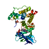
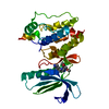
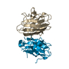
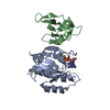
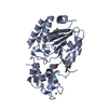
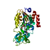
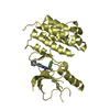
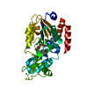
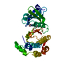
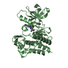
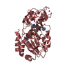
 Homo sapiens (human)
 X-RAY DIFFRACTION /
  Authors
 Citation
 Structure visualization
 Downloads & links
 Other downloads


 PDBj
PDBj Assembly
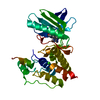

 Mass: 35.453 Da / Num. of mol.: 1 / Source method: obtained synthetically / Formula: Cl
 Mass: 389.473 Da / Num. of mol.: 1 / Source method: obtained synthetically / Formula: C21H19N5OS
 Mass: 18.015 Da / Num. of mol.: 33 / Source method: isolated from a natural source / Formula: H2O
 Sample preparation
 Processing