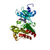
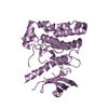
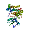
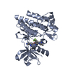
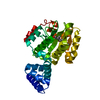
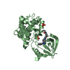
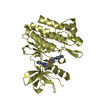
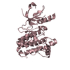
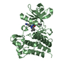
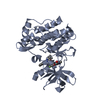
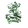
Entry Database : PDB / ID : 3qrjTitle The crystal structure of human abl1 kinase domain T315I mutant in complex with DCC-2036 Tyrosine-protein kinase ABL1 Keywords / / / / / / Function / homology Function Domain/homology Component
/ / / / / / / / / / / / / / / / / / / / / / / / / / / / / / / / / / / / / / / / / / / / / / / / / / / / / / / / / / / / / / / / / / / / / / / / / / / / / / / / / / / / / / / / / / / / / / / / / / / / / / / / / / / / / / / / / / / / / / / / / / / / / / / / / / / / / / / / / / / / / / / / / / / / / / Biological species Homo sapiens (human)Method / / / Resolution : 1.82 Å Authors Chan, W.W. / Wise, S.C. / Kaufman, M.D. / Ahn, Y.M. / Ensinger, C.L. / Haack, T. / Hood, M.M. / Jones, J. / Lord, J.W. / Lu, W.P. ...Chan, W.W. / Wise, S.C. / Kaufman, M.D. / Ahn, Y.M. / Ensinger, C.L. / Haack, T. / Hood, M.M. / Jones, J. / Lord, J.W. / Lu, W.P. / Miller, D. / Patt, W.C. / Smith, B.D. / Petillo, P.A. / Rutkoski, T.J. / Telikepalli, H. / Vogeti, L. / Yao, T. / Chun, L. / Clark, R. / Evangelista, P. / Gavrilescu, L.C. / Lazarides, K. / Zaleskas, V.M. / Stewart, L.J. / Van Etten, R.A. / Flynn, D.L. Journal : Cancer Cell / Year : 2011Title : Conformational Control Inhibition of the BCR-ABL1 Tyrosine Kinase, Including the Gatekeeper T315I Mutant, by the Switch-Control Inhibitor DCC-2036.Authors: Chan, W.W. / Wise, S.C. / Kaufman, M.D. / Ahn, Y.M. / Ensinger, C.L. / Haack, T. / Hood, M.M. / Jones, J. / Lord, J.W. / Lu, W.P. / Miller, D. / Patt, W.C. / Smith, B.D. / Petillo, P.A. / ... Authors : Chan, W.W. / Wise, S.C. / Kaufman, M.D. / Ahn, Y.M. / Ensinger, C.L. / Haack, T. / Hood, M.M. / Jones, J. / Lord, J.W. / Lu, W.P. / Miller, D. / Patt, W.C. / Smith, B.D. / Petillo, P.A. / Rutkoski, T.J. / Telikepalli, H. / Vogeti, L. / Yao, T. / Chun, L. / Clark, R. / Evangelista, P. / Gavrilescu, L.C. / Lazarides, K. / Zaleskas, V.M. / Stewart, L.J. / Van Etten, R.A. / Flynn, D.L. History Deposition Feb 18, 2011 Deposition site / Processing site Revision 1.0 Jun 1, 2011 Provider / Type Revision 1.1 Jul 13, 2011 Group Revision 1.2 Feb 21, 2024 Group / Database references / Derived calculationsCategory chem_comp_atom / chem_comp_bond ... chem_comp_atom / chem_comp_bond / database_2 / struct_ref_seq_dif / struct_site Item _database_2.pdbx_DOI / _database_2.pdbx_database_accession ... _database_2.pdbx_DOI / _database_2.pdbx_database_accession / _struct_ref_seq_dif.details / _struct_site.pdbx_auth_asym_id / _struct_site.pdbx_auth_comp_id / _struct_site.pdbx_auth_seq_id Revision 1.3 Apr 3, 2024 Group / Category
Show all Show less
 Movie
Movie Controller

 Yorodumi
 Open data
 Basic information
 Components
 Keywords
 Function and homology information
 Homo sapiens (human)
 X-RAY DIFFRACTION /
  Authors
 Citation
 Structure visualization
 Downloads & links
 Other downloads
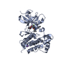


 PDBj
PDBj


 Assembly
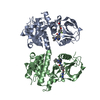


 SPODOPTERA FRUGIPERDA (fall armyworm)
 Mass: 553.587 Da / Num. of mol.: 2 / Source method: obtained synthetically / Formula: C30H28FN7O3
 Mass: 18.015 Da / Num. of mol.: 243 / Source method: isolated from a natural source / Formula: H2O
 Sample preparation
 / Beamline: 5.0.2 / Wavelength: 1 / Wavelength: 1 Å
 Processing