- PDB-1o6y: Catalytic domain of PknB kinase from Mycobacterium tuberculosis -
+
Open data
ID or keywords:
Loading...
-
Basic information
Entry
Database: PDB / ID: 1o6y
Title
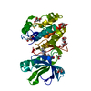
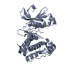
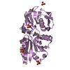
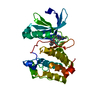
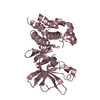
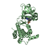
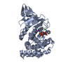
Catalytic domain of PknB kinase from Mycobacterium tuberculosis
Components
SERINE/THREONINE-PROTEIN KINASE PKNB
Keywords
TRANSFERASE / SERINE/THREONINE PROTEIN KINASE / PSI / PROTEIN STRUCTURE INITIATIVE / TB STRUCTURAL GENOMICS CONSORTIUM / TB / TBSGC
Function / homology
Function and homology information
negative regulation of growth rate / negative regulation of fatty acid biosynthetic process / membrane => GO:0016020 / peptidoglycan biosynthetic process / protein serine/threonine/tyrosine kinase activity / peptidoglycan-based cell wall / manganese ion binding / regulation of cell shape / protein kinase activity / non-specific serine/threonine protein kinase ...negative regulation of growth rate / negative regulation of fatty acid biosynthetic process / membrane => GO:0016020 / peptidoglycan biosynthetic process / protein serine/threonine/tyrosine kinase activity / peptidoglycan-based cell wall / manganese ion binding / regulation of cell shape / protein kinase activity / non-specific serine/threonine protein kinase / protein serine kinase activity / protein serine/threonine kinase activity / regulation of DNA-templated transcription / ATP binding / metal ion binding / identical protein binding / plasma membrane Similarity search - Function
PASTA domain / PASTA domain / PASTA domain profile. / PASTA / Phosphorylase Kinase; domain 1 / Phosphorylase Kinase; domain 1 / Transferase(Phosphotransferase) domain 1 / Transferase(Phosphotransferase); domain 1 / Serine/threonine-protein kinase, active site / Serine/Threonine protein kinases active-site signature. ...PASTA domain / PASTA domain / PASTA domain profile. / PASTA / Phosphorylase Kinase; domain 1 / Phosphorylase Kinase; domain 1 / Transferase(Phosphotransferase) domain 1 / Transferase(Phosphotransferase); domain 1 / Serine/threonine-protein kinase, active site / Serine/Threonine protein kinases active-site signature. / Protein kinase domain / Serine/Threonine protein kinases, catalytic domain / Protein kinase, ATP binding site / Protein kinases ATP-binding region signature. / Protein kinase domain profile. / Protein kinase domain / Protein kinase-like domain superfamily / 2-Layer Sandwich / Orthogonal Bundle / Mainly Alpha / Alpha Beta Similarity search - Domain/homology
Mass: 18.015 Da / Num. of mol.: 85 / Source method: isolated from a natural source / Formula: H2O
Sequence details
RESIDUES IN THE N-TERMINAL TAG (1-22) ARE DISORDERED. SIXTEEN RESIDUES (164-179) IN THE ACTIVATION ...RESIDUES IN THE N-TERMINAL TAG (1-22) ARE DISORDERED. SIXTEEN RESIDUES (164-179) IN THE ACTIVATION LOOP ARE ALSO DISORDERED.
-
Experimental details
-
Experiment
Experiment
Method: X-RAY DIFFRACTION / Number of used crystals: 1
-
Sample preparation
Crystal
Density Matthews: 2.22 Å3/Da / Density % sol: 44.17 %
Crystal grow
Temperature: 292 K / Method: vapor diffusion, hanging drop / pH: 7.5 Details: PROTEIN CRYSTALLIZED FROM 27% PEG 400, 4% 1,3-BUTANEDIOL, 100 MM HEPES, PH 7.5, 30 MM MGCL2 AT 19 DEGREES C
Crystal grow
*PLUS
Temperature: 19 ℃ / Method: vapor diffusion, hanging drop
 Movie
Movie Controller


 Open data
 Basic information
 Components
 Keywords
 Function and homology information
 MYCOBACTERIUM TUBERCULOSIS (bacteria)
 X-RAY DIFFRACTION /
  Authors
 Citation
 Structure visualization
 Downloads & links
 Other downloads
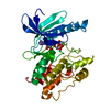


 PDBj
PDBj


 Assembly

 Mass: 505.208 Da / Num. of mol.: 1 / Source method: obtained synthetically / Formula: C11H18N5O12P3 / Comment: AMP-PCP, energy-carrying molecule analogue*YM
 Mass: 24.305 Da / Num. of mol.: 2 / Source method: obtained synthetically / Formula: Mg
 Mass: 18.015 Da / Num. of mol.: 85 / Source method: isolated from a natural source / Formula: H2O
 Sample preparation
 / Beamline: ID14-2 / Wavelength: 0.933
  Processing