Mass: 18.015 Da / Num. of mol.: 529 / Source method: isolated from a natural source / Formula: H2O
Has protein modification
Y
Sequence details
THIS CONSTRUCT (RESIDUES 28-272) WAS EXPRESSED WITH A PURIFICATION TAG MGSDKIHHHHHHENLYFQG. THE TAG ...THIS CONSTRUCT (RESIDUES 28-272) WAS EXPRESSED WITH A PURIFICATION TAG MGSDKIHHHHHHENLYFQG. THE TAG WAS REMOVED WITH TEV PROTEASE LEAVING ONLY A GLYCINE (0) FOLLOWED BY THE TARGET SEQUENCE.
-
Experimental details
-
Experiment
Experiment
Method: X-RAY DIFFRACTION / Number of used crystals: 1
-
Sample preparation
Crystal
Density Matthews: 2.38 Å3/Da / Density % sol: 48.31 % Description: THIS STRUCTURE WAS SOLVED BY MOLECULAR REPLACEMENT WITH PHASER USING AS A MODEL THE STRUCTURE OF THE SAME PROTEIN SOLVED BY MAD PHASING AT 2.10 A IN SPACE GROUP P212121
Crystal grow
Temperature: 277 K / Method: vapor diffusion, sitting drop / pH: 5.6 Details: 20.0% iso-Propanol, 20.0% PEG-4000, 0.1M Citrate pH 5.6, NANODROP, VAPOR DIFFUSION, SITTING DROP, temperature 277K
Type: DECTRIS PILATUS 6M / Detector: PIXEL / Date: Feb 8, 2012 Details: Flat mirror (vertical focusing); single crystal Si(111) bent monochromator (horizontal focusing)
Radiation
Monochromator: single crystal Si(111) bent / Protocol: SINGLE WAVELENGTH / Monochromatic (M) / Laue (L): M / Scattering type: x-ray
Radiation wavelength
Wavelength: 0.97908 Å / Relative weight: 1
Reflection
Resolution: 1.9→29.38 Å / Num. obs: 39597 / % possible obs: 92.4 % / Observed criterion σ(I): -3 / Biso Wilson estimate: 18.401 Å2 / Rmerge(I) obs: 0.062 / Net I/σ(I): 10.04
Reflection shell
Diffraction-ID: 1
Resolution (Å)
Highest resolution (Å)
Rmerge(I) obs
Mean I/σ(I) obs
Num. measured obs
Num. unique obs
% possible all
1.9-1.97
0.353
2.3
13977
7523
91.8
1.97-2.05
0.257
3.2
13960
7428
92.2
2.05-2.14
0.208
3.9
13275
7103
92.8
2.14-2.25
0.164
4.7
13228
7167
91.8
2.25-2.39
0.134
5.6
12465
7185
90
2.39-2.58
0.094
7.8
14413
7676
93.6
2.58-2.84
0.067
10.8
14195
7539
94
2.84-3.25
0.044
15.3
13555
7423
93.5
3.25-4.08
0.029
21.1
12906
7245
91.5
4.08
0.024
25.1
13805
7503
92.8
-
Phasing
Phasing
Method: molecular replacement
-
Processing
Software
Name
Version
Classification
NB
MolProbity
3beta29
modelbuilding
PDB_EXTRACT
3.1
dataextraction
PHASER
2.3.0
phasing
XSCALE
December29, 2011
datascaling
REFMAC
5.6.0117
refinement
XDS
datareduction
Refinement
Method to determine structure: MOLECULAR REPLACEMENT / Resolution: 1.9→29.38 Å / Cor.coef. Fo:Fc: 0.946 / Cor.coef. Fo:Fc free: 0.916 / Occupancy max: 1 / Occupancy min: 0.5 / SU B: 7.647 / SU ML: 0.12 / Cross valid method: THROUGHOUT / σ(F): 0 / ESU R: 0.175 / ESU R Free: 0.16 Stereochemistry target values: MAXIMUM LIKELIHOOD WITH PHASES Details: 1.HYDROGENS HAVE BEEN ADDED IN THE RIDING POSITIONS. 2.A MET-INHIBITION PROTOCOL WAS USED FOR SELENOMETHIONINE INCORPORATION DURING PROTEIN EXPRESSION. THE OCCUPANCY OF THE SE ATOMS IN THE ...Details: 1.HYDROGENS HAVE BEEN ADDED IN THE RIDING POSITIONS. 2.A MET-INHIBITION PROTOCOL WAS USED FOR SELENOMETHIONINE INCORPORATION DURING PROTEIN EXPRESSION. THE OCCUPANCY OF THE SE ATOMS IN THE MSE RESIDUES WAS REDUCED TO 0.75 FOR THE REDUCED SCATTERING POWER DUE TO PARTIAL S-MET INCORPORATION. 3.ATOM RECORD CONTAINS SUM OF TLS AND RESIDUAL B FACTORS. ANISOU RECORD CONTAINS SUM OF TLS AND RESIDUAL U FACTORS. 4.WATERS WERE EXCLUDED FROM AUTOMATIC TLS ASSIGNMENT. 5.SELENO-METHIONINE (MSE) HAS BEEN MODELED AT THE PUTATIVE ACTIVE SITE BASED ON PUTATIVE ASSIGNMENT OF FUNCTION FROM PROTEIN STRUCTURE COMPARISONS AND ANOMALOUS DIFFERENCE FOURIER ELECTRON DENSITY MAPS. 6.RAMACHANDRAN OUTLIERS AT PHE84 IN BOTH PROTEIN ARE SUPPORTED BY ELECTRON DENSITY AND LIE IN THE VICINITY OF THE PUTATIVE ACTIVE SITE. 7. NCS RESTRAINTS WERE APPLIED USING REFMAC's LOCAL NCS OPTION (NCSR LOCAL). NCS GROUP 1 CHAIN A (28-272 TO CHAIN B (28-272) COUNT: 8927, RMS: 0.09, WEIGHT: 0.05.
Rfactor
Num. reflection
% reflection
Selection details
Rfree
0.2382
1990
5 %
RANDOM
Rwork
0.1884
-
-
-
obs
0.1909
39575
97.76 %
-
Solvent computation
Ion probe radii: 0.8 Å / Shrinkage radii: 0.8 Å / VDW probe radii: 1.2 Å / Solvent model: BABINET MODEL WITH MASK
In the structure databanks used in Yorodumi, some data are registered as the other names, "COVID-19 virus" and "2019-nCoV". Here are the details of the virus and the list of structure data.
Jan 31, 2019. EMDB accession codes are about to change! (news from PDBe EMDB page)
EMDB accession codes are about to change! (news from PDBe EMDB page)
The allocation of 4 digits for EMDB accession codes will soon come to an end. Whilst these codes will remain in use, new EMDB accession codes will include an additional digit and will expand incrementally as the available range of codes is exhausted. The current 4-digit format prefixed with “EMD-” (i.e. EMD-XXXX) will advance to a 5-digit format (i.e. EMD-XXXXX), and so on. It is currently estimated that the 4-digit codes will be depleted around Spring 2019, at which point the 5-digit format will come into force.
The EM Navigator/Yorodumi systems omit the EMD- prefix.
Related info.:Q: What is EMD? / ID/Accession-code notation in Yorodumi/EM Navigator
Yorodumi is a browser for structure data from EMDB, PDB, SASBDB, etc.
This page is also the successor to EM Navigator detail page, and also detail information page/front-end page for Omokage search.
The word "yorodu" (or yorozu) is an old Japanese word meaning "ten thousand". "mi" (miru) is to see.
Related info.:EMDB / PDB / SASBDB / Comparison of 3 databanks / Yorodumi Search / Aug 31, 2016. New EM Navigator & Yorodumi / Yorodumi Papers / Jmol/JSmol / Function and homology information / Changes in new EM Navigator and Yorodumi
 Movie
Movie Controller

 Yorodumi
 Open data
 Basic information
 Components
 Keywords
 Function and homology information
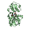
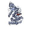
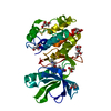
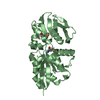
 Enterococcus faecalis (bacteria)
 X-RAY DIFFRACTION /
  Authors
 Citation
 Structure visualization
 Downloads & links
 Other downloads
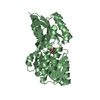


 PDBj
PDBj


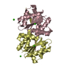
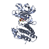
 Assembly


 Type: L-peptide linking / Mass: 196.106 Da / Num. of mol.: 2 / Source method: obtained synthetically / Formula: C5H11NO2Se
 Mass: 18.015 Da / Num. of mol.: 529 / Source method: isolated from a natural source / Formula: H2O
 Sample preparation
 / Beamline: BL11-1 / Wavelength: 0.97908
  Processing