 Movie
Movie Controller

[English] 日本語
 Yorodumi
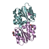
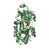
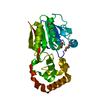
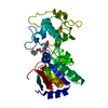
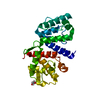
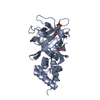
- PDB-3ipr: Crystal structure of the Enterococcus faecalis gluconate specific... -
+ Open data

- Basic information
| Entry | Database: PDB / ID: 3ipr | ||||||
|---|---|---|---|---|---|---|---|
| Title | Crystal structure of the Enterococcus faecalis gluconate specific EIIA phosphotransferase system component | ||||||
 Components Components | PTS system, IIA component | ||||||
 Keywords Keywords | TRANSFERASE / 4 stranded parallel BETA-SHEET flanked by 3 ALPHA-HELICES on each side | ||||||
| Function / homology |  Function and homology information Function and homology informationphosphoenolpyruvate-dependent sugar phosphotransferase system / kinase activity / membrane / metal ion binding / cytoplasm Similarity search - Function | ||||||
| Biological species |   Enterococcus faecalis (bacteria) Enterococcus faecalis (bacteria) | ||||||
| Method |  X-RAY DIFFRACTION / SYNCHROTRON / MOLECULAR REPLACEMENT / molecular replacement / Resolution: 2.5 Å X-RAY DIFFRACTION / SYNCHROTRON / MOLECULAR REPLACEMENT / molecular replacement / Resolution: 2.5 Å | ||||||
 Authors Authors | Reinelt, S. / Welti, S. / Scheffzek, K. | ||||||
 Citation Citation | Journal: Biochem.Biophys.Res.Commun. / Year: 2009 Title: Structure of the Enterococcus faecalis EIIA(gnt) PTS component. Authors: Reinelt, S. / Koch, B. / Hothorn, M. / Hengstenberg, W. / Welti, S. / Scheffzek, K. | ||||||
| History |
|
- Structure visualization
| Structure viewer | Molecule: MolmilJmol/JSmol |
|---|
- Downloads & links
-Download
| PDBx/mmCIF format | 3ipr.cif.gz | 159.8 KB | Display | PDBx/mmCIF format |
|---|---|---|---|---|
| PDB format | pdb3ipr.ent.gz | 128.4 KB | Display | PDB format |
| PDBx/mmJSON format | 3ipr.json.gz | Tree view | PDBx/mmJSON format | |
| Others |  Other downloads Other downloads |
-Validation report
| Arichive directory | https://data.pdbj.org/pub/pdb/validation_reports/ip/3iprftp://data.pdbj.org/pub/pdb/validation_reports/ip/3ipr | HTTPS FTP |
|---|
-Related structure data
| Related structure data | 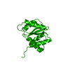 1pdoS S: Starting model for refinement |
|---|---|
| Similar structure data |


-Links
 PDBj
PDBj- Assembly
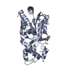
| Deposited unit | 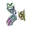
| ||||||||
|---|---|---|---|---|---|---|---|---|---|
| 1 | 
| ||||||||
| 2 | 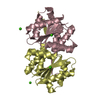
| ||||||||
| 3 | 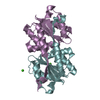
| ||||||||
| Unit cell |
|
-Components
| #1: Protein | Mass: 15798.622 Da / Num. of mol.: 6 Source method: isolated from a genetically manipulated source Details: gluconate specific EIIA phosphotransferase system component from Enterococcus faecalis Source: (gene. exp.) Enterococcus faecalis (bacteria) / Strain: 26487 Wildtype / Gene: EF_3136, gntF / Plasmid: pET11a / Production host: #2: Chemical | ChemComp-CA /   Mass: 40.078 Da / Num. of mol.: 10 / Source method: obtained synthetically / Formula: Ca Mass: 40.078 Da / Num. of mol.: 10 / Source method: obtained synthetically / Formula: Ca#3: Water | ChemComp-HOH / |  Mass: 18.015 Da / Num. of mol.: 117 / Source method: isolated from a natural source / Formula: H2O Mass: 18.015 Da / Num. of mol.: 117 / Source method: isolated from a natural source / Formula: H2O |
|---|
-Experimental details
-Experiment
| Experiment | Method: X-RAY DIFFRACTION / Number of used crystals: 1 |
|---|
- Sample preparation
| Crystal | Density Matthews: 2.47 Å3/Da / Density % sol: 50.26 % |
|---|---|
| Crystal grow | Temperature: 291.15 K / Method: vapor diffusion, hanging drop / pH: 6.5 Details: 18% PEG 4000, 0.2M Ca acetate, 0.1M Ca cacodylate, pH 6.5, VAPOR DIFFUSION, HANGING DROP, temperature 291.15K |
-Data collection
| Diffraction | Mean temperature: 100 K |
|---|---|
| Diffraction source | Source: SYNCHROTRON / Site: ESRF  / Beamline: ID14-1 / Wavelength: 1.044 Å / Beamline: ID14-1 / Wavelength: 1.044 Å |
| Detector | Type: ADSC QUANTUM 210 / Detector: CCD / Date: Dec 11, 2003 / Details: mirrors |
| Radiation | Monochromator: Diamond(111) / Protocol: SINGLE WAVELENGTH / Monochromatic (M) / Laue (L): M / Scattering type: x-ray |
| Radiation wavelength | Wavelength: 1.044 Å / Relative weight: 1 |
| Reflection | Resolution: 2.5→20 Å / Num. all: 30490 / Num. obs: 30490 / % possible obs: 94.4 % / Observed criterion σ(I): 0 / Redundancy: 3.7 % |
| Reflection shell | Resolution: 2.5→2.6 Å / Mean I/σ(I) obs: 4.42 / % possible all: 94.3 |
-Phasing
| Phasing | Method: molecular replacement |
|---|
- Processing
| Software |
| |||||||||||||||||||||||||
|---|---|---|---|---|---|---|---|---|---|---|---|---|---|---|---|---|---|---|---|---|---|---|---|---|---|---|
| Refinement | Method to determine structure: MOLECULAR REPLACEMENT Starting model: PDB entry 1PDO Resolution: 2.5→20 Å / Occupancy max: 1 / Occupancy min: 1 / Cross valid method: FREE R / σ(F): 0 / Stereochemistry target values: Engh & Huber
| |||||||||||||||||||||||||
| Solvent computation | Bsol: 45.951 Å2 | |||||||||||||||||||||||||
| Displacement parameters | Biso max: 104.14 Å2 / Biso mean: 33.949 Å2 / Biso min: 3.17 Å2
| |||||||||||||||||||||||||
| Refinement step | Cycle: LAST / Resolution: 2.5→20 Å
| |||||||||||||||||||||||||
| Refine LS restraints |
| |||||||||||||||||||||||||
| Xplor file |
|
