 Movie
Movie Controller


+ Open data

- Basic information
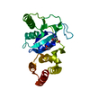
| Entry | Database: PDB / ID: 1pdo | ||||||
|---|---|---|---|---|---|---|---|
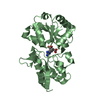
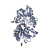
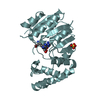
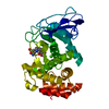
| Title | PHOSPHOENOLPYRUVATE-DEPENDENT PHOSPHOTRANSFERASE SYSTEM | ||||||
 Components Components | MANNOSE PERMEASE | ||||||
 Keywords Keywords | PHOSPHOTRANSFERASE / PHOSPHOENOLPYRUVATE DEPENDENT PHOSPHOTRANSFERASE SYSTEM | ||||||
| Function / homology |  Function and homology information Function and homology informationprotein-Npi-phosphohistidine-D-mannose phosphotransferase / mannose transmembrane transport / protein-N(PI)-phosphohistidine-mannose phosphotransferase system transporter activity / fructose import across plasma membrane / N-acetylglucosamine transport / protein-N(PI)-phosphohistidine-carbohydrate phosphotransferase activity / D-glucose import across plasma membrane / phosphoenolpyruvate-dependent sugar phosphotransferase system / transmembrane transporter complex / kinase activity ...protein-Npi-phosphohistidine-D-mannose phosphotransferase / mannose transmembrane transport / protein-N(PI)-phosphohistidine-mannose phosphotransferase system transporter activity / fructose import across plasma membrane / N-acetylglucosamine transport / protein-N(PI)-phosphohistidine-carbohydrate phosphotransferase activity / D-glucose import across plasma membrane / phosphoenolpyruvate-dependent sugar phosphotransferase system / transmembrane transporter complex / kinase activity / protein homodimerization activity / membrane / plasma membrane / cytoplasm / cytosol Similarity search - Function | ||||||
| Biological species |  | ||||||
| Method |  X-RAY DIFFRACTION / SYNCHROTRON / MIRAS SOFTWARE USED : CCP4 PROGRAM SUITE 1994 STARTING MODEL FOR MOLECULAR REPLACEMENT: NULL / Resolution: 1.7 Å X-RAY DIFFRACTION / SYNCHROTRON / MIRAS SOFTWARE USED : CCP4 PROGRAM SUITE 1994 STARTING MODEL FOR MOLECULAR REPLACEMENT: NULL / Resolution: 1.7 Å | ||||||
 Authors Authors | Nunn, R.S. / Erni, B. / Schirmer, T. | ||||||
 Citation Citation | Journal: J.Mol.Biol. / Year: 1996 Title: Structure of the IIA domain of the mannose transporter from Escherichia coli at 1.7 angstroms resolution. Authors: Nunn, R.S. / Markovic-Housley, Z. / Genovesio-Taverne, J.C. / Flukiger, K. / Rizkallah, P.J. / Jansonius, J.N. / Schirmer, T. / Erni, B. #1: Journal: J.Biol.Chem. / Year: 1987Title: The Mannose Permease of Escherichia Coli Consists of Three Different Proteins. Amino Acid Sequence and Function in Sugar Transport, Sugar Phosphorylation, and Penetration of Phage Lambda DNA Authors: Erni, B. / Zanolari, B. / Kocher, H.P. | ||||||
| History |
|
- Structure visualization
| Structure viewer | Molecule: MolmilJmol/JSmol |
|---|
- Downloads & links
-Download
| PDBx/mmCIF format | 1pdo.cif.gz | 38.2 KB | Display | PDBx/mmCIF format |
|---|---|---|---|---|
| PDB format | pdb1pdo.ent.gz | 26.6 KB | Display | PDB format |
| PDBx/mmJSON format | 1pdo.json.gz | Tree view | PDBx/mmJSON format | |
| Others |  Other downloads Other downloads |
-Validation report
| Arichive directory | https://data.pdbj.org/pub/pdb/validation_reports/pd/1pdoftp://data.pdbj.org/pub/pdb/validation_reports/pd/1pdo | HTTPS FTP |
|---|
-Related structure data
| Similar structure data |
|---|


-Links
 PDBj
PDBj- Assembly
| Deposited unit | 
| ||||||||
|---|---|---|---|---|---|---|---|---|---|
| 1 | 
| ||||||||
| Unit cell |
| ||||||||
| Components on special symmetry positions |
|
-Components
| #1: Protein | Mass: 14624.625 Da / Num. of mol.: 1 Fragment: IIA ==MAN== DOMAIN, RESIDUES 2 - 133, OF THE IIAB ==MAN== SUBUNIT PLUS PHE-ALA-GLY AT THE CARBOXY TERMINUS Source method: isolated from a genetically manipulated source Source: (gene. exp.) Description: SUBCLONAL IIA COMPRISES RESIDUES 1 - 133 OF THE WILD-TYPE IIAB SEQUENCE PLUS PHE-ALA-GLY AT THE CARBOXY TERMINUS. THE INITIAL SE-MET WAS REMOVED BY PROTEOLYTIC CLEAVAGE. Plasmid: PJFL1320 / Species (production host): Escherichia coli / Gene (production host): MANX / Production host: References: UniProt: P69797, protein-Npi-phosphohistidine-sugar phosphotransferase |
|---|---|
| #2: Water | ChemComp-HOH /  Mass: 18.015 Da / Num. of mol.: 70 / Source method: isolated from a natural source / Formula: H2O Mass: 18.015 Da / Num. of mol.: 70 / Source method: isolated from a natural source / Formula: H2O |
| Compound details | DURING CATALYSIS HIS 10 GETS TRANSIENTL |
-Experimental details
-Experiment
| Experiment | Method: X-RAY DIFFRACTION / Number of used crystals: 3 |
|---|
- Sample preparation
| Crystal | Density Matthews: 2.55 Å3/Da / Density % sol: 46 % | |||||||||||||||||||||||||||||||||||
|---|---|---|---|---|---|---|---|---|---|---|---|---|---|---|---|---|---|---|---|---|---|---|---|---|---|---|---|---|---|---|---|---|---|---|---|---|
| Crystal grow | pH: 6.8 / Details: pH 6.8 | |||||||||||||||||||||||||||||||||||
| Crystal grow | *PLUS Method: vapor diffusion | |||||||||||||||||||||||||||||||||||
| Components of the solutions | *PLUS
|
-Data collection
| Diffraction | Mean temperature: 277 K |
|---|---|
| Diffraction source | Source: SYNCHROTRON / Site: SRS  / Beamline: PX9.6 / Wavelength: 0.87 / Beamline: PX9.6 / Wavelength: 0.87 |
| Detector | Type: MARRESEARCH / Detector: IMAGE PLATE / Date: Sep 3, 1995 |
| Radiation | Monochromatic (M) / Laue (L): M / Scattering type: x-ray |
| Radiation wavelength | Wavelength: 0.87 Å / Relative weight: 1 |
| Reflection | Resolution: 1.7→19.8 Å / Num. obs: 16839 / % possible obs: 97.3 % / Observed criterion σ(I): 4 / Redundancy: 5.9 % / Rmerge(I) obs: 0.068 / Net I/σ(I): 32.4 |
| Reflection shell | Resolution: 1.7→1.74 Å / Redundancy: 5.6 % / Rmerge(I) obs: 0.272 / Mean I/σ(I) obs: 8.1 / % possible all: 95.8 |
| Reflection shell | *PLUS % possible obs: 95.8 % |
- Processing
| Software |
| ||||||||||||||||||||||||||||||||||||||||||||||||||||||||||||||||||||||||||||||||
|---|---|---|---|---|---|---|---|---|---|---|---|---|---|---|---|---|---|---|---|---|---|---|---|---|---|---|---|---|---|---|---|---|---|---|---|---|---|---|---|---|---|---|---|---|---|---|---|---|---|---|---|---|---|---|---|---|---|---|---|---|---|---|---|---|---|---|---|---|---|---|---|---|---|---|---|---|---|---|---|---|---|
| Refinement | Method to determine structure: MIRAS SOFTWARE USED : CCP4 PROGRAM SUITE 1994 STARTING MODEL FOR MOLECULAR REPLACEMENT: NULL Resolution: 1.7→8 Å / σ(F): 0
| ||||||||||||||||||||||||||||||||||||||||||||||||||||||||||||||||||||||||||||||||
| Displacement parameters | Biso mean: 20.2 Å2 | ||||||||||||||||||||||||||||||||||||||||||||||||||||||||||||||||||||||||||||||||
| Refinement step | Cycle: LAST / Resolution: 1.7→8 Å
| ||||||||||||||||||||||||||||||||||||||||||||||||||||||||||||||||||||||||||||||||
| Refine LS restraints |
| ||||||||||||||||||||||||||||||||||||||||||||||||||||||||||||||||||||||||||||||||
| Software | *PLUS Name: X-PLOR / Version: 3.1 / Classification: refinement | ||||||||||||||||||||||||||||||||||||||||||||||||||||||||||||||||||||||||||||||||
| Refine LS restraints | *PLUS
|
