 Movie
Movie Controller

[English] 日本語
 Yorodumi
- PDB-2ore: Binary Structure of Escherichia coli DNA Adenine Methyltransferas... -
+ Open data

- Basic information
| Entry | Database: PDB / ID: 2ore | ||||||
|---|---|---|---|---|---|---|---|
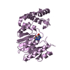
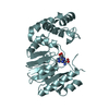
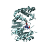
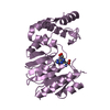
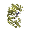
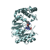
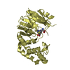
| Title | Binary Structure of Escherichia coli DNA Adenine Methyltransferase and S-adenosylhomocysteine | ||||||
 Components Components | DNA adenine methylase | ||||||
 Keywords Keywords | TRANSFERASE / DAM METHYLATION / GATC RECOGNITION / S-ADENOSYLHOMOCYSTEINE CONFORMATION / BACTERIAL VIRULENCE FACTOR | ||||||
| Function / homology |  Function and homology information Function and homology informationbacterial-type DNA replication initiation / site-specific DNA-methyltransferase (adenine-specific) / site-specific DNA-methyltransferase (adenine-specific) activity / S-adenosyl-L-methionine binding / DNA restriction-modification system / response to UV / mismatch repair / DNA-templated DNA replication / methylation / sequence-specific DNA binding Similarity search - Function | ||||||
| Biological species |  | ||||||
| Method |  X-RAY DIFFRACTION / SYNCHROTRON / MOLECULAR REPLACEMENT / Resolution: 2.99 Å X-RAY DIFFRACTION / SYNCHROTRON / MOLECULAR REPLACEMENT / Resolution: 2.99 Å | ||||||
 Authors Authors | Horton, J.R. / Cheng, X. | ||||||
 Citation Citation | Journal: J.Biol.Chem. / Year: 2007 Title: Two Alternative Conformations of S-Adenosyl-L-homocysteine Bound to Escherichia coli DNA Adenine Methyltransferase and the Implication of Conformational Changes in Regulating the Catalytic Cycle. Authors: Liebert, K. / Horton, J.R. / Chahar, S. / Orwick, M. / Cheng, X. / Jeltsch, A. #1: Journal: J.Mol.Biol. / Year: 2006Title: Structure and substrate recognition of the Escherichia coli DNA adenine methyltransferase. Authors: Horton, J.R. / Liebert, K. / Bekes, M. / Jeltsch, A. / Cheng, X. | ||||||
| History |
|
- Structure visualization
| Structure viewer | Molecule: MolmilJmol/JSmol |
|---|
- Downloads & links
-Download
| PDBx/mmCIF format | 2ore.cif.gz | 161.4 KB | Display | PDBx/mmCIF format |
|---|---|---|---|---|
| PDB format | pdb2ore.ent.gz | 126.5 KB | Display | PDB format |
| PDBx/mmJSON format | 2ore.json.gz | Tree view | PDBx/mmJSON format | |
| Others |  Other downloads Other downloads |
-Validation report
| Arichive directory | https://data.pdbj.org/pub/pdb/validation_reports/or/2oreftp://data.pdbj.org/pub/pdb/validation_reports/or/2ore | HTTPS FTP |
|---|
-Related structure data
| Related structure data | 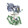 2g1pS S: Starting model for refinement |
|---|---|
| Similar structure data |


-Links
 PDBj
PDBj- Assembly
| Deposited unit | 
| ||||||||
|---|---|---|---|---|---|---|---|---|---|
| 1 | 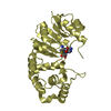
| ||||||||
| 2 | 
| ||||||||
| 3 | 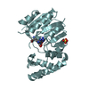
| ||||||||
| Unit cell |
|
-Components
| #1: Protein | Mass: 34314.961 Da / Num. of mol.: 3 Source method: isolated from a genetically manipulated source Source: (gene. exp.) References: UniProt: P0AEE8, site-specific DNA-methyltransferase (adenine-specific) #2: Chemical |   Type: L-peptide linking / Mass: 384.411 Da / Num. of mol.: 3 / Source method: obtained synthetically / Formula: C14H20N6O5S Type: L-peptide linking / Mass: 384.411 Da / Num. of mol.: 3 / Source method: obtained synthetically / Formula: C14H20N6O5S#3: Chemical | ChemComp-SO4 / |   Mass: 96.063 Da / Num. of mol.: 1 / Source method: obtained synthetically / Formula: SO4 Mass: 96.063 Da / Num. of mol.: 1 / Source method: obtained synthetically / Formula: SO4#4: Water | ChemComp-HOH / |  Mass: 18.015 Da / Num. of mol.: 23 / Source method: isolated from a natural source / Formula: H2O Mass: 18.015 Da / Num. of mol.: 23 / Source method: isolated from a natural source / Formula: H2O |
|---|
-Experimental details
-Experiment
| Experiment | Method: X-RAY DIFFRACTION / Number of used crystals: 1 |
|---|
- Sample preparation
| Crystal | Density Matthews: 3.61 Å3/Da / Density % sol: 65.9 % |
|---|---|
| Crystal grow | Temperature: 289 K / Method: vapor diffusion, hanging drop / pH: 7.2 Details: 1.5 M lithium sulfate,50mM magnesium sulfate, 100mM HEPES pH 7.2, VAPOR DIFFUSION, HANGING DROP, temperature 289K |
-Data collection
| Diffraction | Mean temperature: 173 K |
|---|---|
| Diffraction source | Source: SYNCHROTRON / Site: APS  / Beamline: 22-ID / Wavelength: 0.97923 Å / Beamline: 22-ID / Wavelength: 0.97923 Å |
| Detector | Type: MARRESEARCH / Detector: CCD / Date: Apr 18, 2005 |
| Radiation | Monochromator: Si 111 CHANNEL / Protocol: SINGLE WAVELENGTH / Monochromatic (M) / Laue (L): M / Scattering type: x-ray |
| Radiation wavelength | Wavelength: 0.97923 Å / Relative weight: 1 |
| Reflection | Resolution: 2.99→34.67 Å / Num. all: 28266 / Num. obs: 28125 / % possible obs: 99.5 % / Observed criterion σ(F): -3 / Observed criterion σ(I): -3 / Rmerge(I) obs: 0.111 |
| Reflection shell | Resolution: 2.99→3.1 Å / % possible all: 100 |
- Processing
| Software |
| |||||||||||||||||||||||||
|---|---|---|---|---|---|---|---|---|---|---|---|---|---|---|---|---|---|---|---|---|---|---|---|---|---|---|
| Refinement | Method to determine structure: MOLECULAR REPLACEMENT Starting model: 2G1P Resolution: 2.99→34.67 Å / Isotropic thermal model: ISOTROPIC / Cross valid method: THROUGHOUT / σ(F): -3 / σ(I): -3 / Stereochemistry target values: Engh & Huber
| |||||||||||||||||||||||||
| Displacement parameters | Biso mean: 38.9 Å2
| |||||||||||||||||||||||||
| Refine analyze |
| |||||||||||||||||||||||||
| Refinement step | Cycle: LAST / Resolution: 2.99→34.67 Å
| |||||||||||||||||||||||||
| Refine LS restraints |
|
