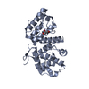
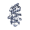
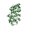
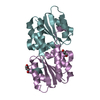
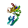
[English] 日本語
Yorodumi
- PDB-6oa6: CRYSTAL STRUCTURE OF THE ACTIN-BINDING DOMAIN OF HUMAN ALPHA-ACTININ-4 -

+
Open data


ID or keywords:

Loading...

-
Basic information

Entry
Database: PDB / ID: 6oa6
TitleCRYSTAL STRUCTURE OF THE ACTIN-BINDING DOMAIN OF HUMAN ALPHA-ACTININ-4
ComponentsAlpha-actinin-4
KeywordsSTRUCTURAL PROTEIN / CALPONIN HOMOLOGY DOMAIN / CH DOMAIN / ACTIN- BINDING PROTEIN / ACTIN-CROSSLINKING / GLOMERULOSCLEROS SPECTRIN FAMILY / DISEASE MUTATION / NUCLEUS / PHOSPHORYLATION
Function / homology
Function and homology information


positive regulation of sodium:proton antiporter activity / negative regulation of substrate adhesion-dependent cell spreading / nucleoside binding / muscle cell development / nuclear retinoic acid receptor binding / vesicle transport along actin filament / Nephrin family interactions / cortical actin cytoskeleton / pseudopodium / retinoic acid receptor signaling pathway ...positive regulation of sodium:proton antiporter activity / negative regulation of substrate adhesion-dependent cell spreading / nucleoside binding / muscle cell development / nuclear retinoic acid receptor binding / vesicle transport along actin filament / Nephrin family interactions / cortical actin cytoskeleton / pseudopodium / retinoic acid receptor signaling pathway / stress fiber / tumor necrosis factor-mediated signaling pathway / peroxisome proliferator activated receptor signaling pathway / nuclear receptor coactivator activity / platelet alpha granule lumen / cell projection / nuclear receptor binding / RNA polymerase II transcription regulatory region sequence-specific DNA binding / chromatin DNA binding / Z disc / positive regulation of non-canonical NF-kappaB signal transduction / actin filament binding / actin cytoskeleton / protein transport / integrin binding / Platelet degranulation / cell junction / actin binding / actin cytoskeleton organization / regulation of apoptotic process / transmembrane transporter binding / transcription coactivator activity / positive regulation of cell migration / ribonucleoprotein complex / focal adhesion / calcium ion binding / perinuclear region of cytoplasm / protein homodimerization activity / positive regulation of transcription by RNA polymerase II / protein-containing complex / extracellular space / RNA binding / extracellular exosome / extracellular region / nucleus / plasma membrane / cytoplasm
Similarity search - Function
Calponin-like domain / EF-hand, Ca insensitive / Ca2+ insensitive EF hand / Ca2+ insensitive EF hand / Actin-binding Protein, T-fimbrin; domain 1 / Spectrin repeat / Spectrin repeat / Actinin-type actin-binding domain signature 1. / Actinin-type actin-binding domain signature 2. / Actinin-type actin-binding domain, conserved site ...Calponin-like domain / EF-hand, Ca insensitive / Ca2+ insensitive EF hand / Ca2+ insensitive EF hand / Actin-binding Protein, T-fimbrin; domain 1 / Spectrin repeat / Spectrin repeat / Actinin-type actin-binding domain signature 1. / Actinin-type actin-binding domain signature 2. / Actinin-type actin-binding domain, conserved site / Spectrin/alpha-actinin / Spectrin repeats / Calponin homology domain / Calponin homology (CH) domain / Calponin homology domain / CH domain superfamily / Calponin homology (CH) domain profile. / EF-hand, calcium binding motif / EF-Hand 1, calcium-binding site / EF-hand calcium-binding domain. / EF-hand calcium-binding domain profile. / EF-hand domain / EF-hand domain pair / Orthogonal Bundle / Mainly Alpha
Similarity search - Domain/homology
Biological speciesHomo sapiens (human)
MethodX-RAY DIFFRACTION / SYNCHROTRON / MOLECULAR REPLACEMENT / Resolution: 1.37 Å
AuthorsBirrane, G. / Feng, D.
Funding support United States, 3items
OrganizationGrant numberCountry
National Institutes of Health/National Institute of Diabetes and Digestive and Kidney Disease (NIH/NIDDK)DK59588 United States
National Institutes of Health/National Institute of Diabetes and Digestive and Kidney Disease (NIH/NIDDK)DK114329 United States
National Institutes of Health/National Institute of Diabetes and Digestive and Kidney Disease (NIH/NIDDK)DK007199 United States
CitationJournal: J. Am. Soc. Nephrol. / Year: 2020
Title: Phosphorylation of ACTN4 Leads to Podocyte Vulnerability and Proteinuric Glomerulosclerosis.
Authors: Feng, D. / Kumar, M. / Muntel, J. / Gurley, S.B. / Birrane, G. / Stillman, I.E. / Ding, L. / Wang, M. / Ahmed, S. / Schlondorff, J. / Alper, S.L. / Ferrante, T. / Marquez, S.L. / Ng, C.F. / ...Authors: Feng, D. / Kumar, M. / Muntel, J. / Gurley, S.B. / Birrane, G. / Stillman, I.E. / Ding, L. / Wang, M. / Ahmed, S. / Schlondorff, J. / Alper, S.L. / Ferrante, T. / Marquez, S.L. / Ng, C.F. / Novak, R. / Ingber, D.E. / Steen, H. / Pollak, M.R.
History
DepositionMar 15, 2019Deposition site: RCSB / Processing site: RCSB
Revision 1.0Mar 18, 2020Provider: repository / Type: Initial release
Revision 1.1Sep 9, 2020Group: Database references / Category: citation / citation_author
Item: _citation.journal_abbrev / _citation.journal_id_CSD ..._citation.journal_abbrev / _citation.journal_id_CSD / _citation.journal_id_ISSN / _citation.journal_volume / _citation.page_first / _citation.page_last / _citation.pdbx_database_id_DOI / _citation.pdbx_database_id_PubMed / _citation.title / _citation.year
Revision 1.2Oct 11, 2023Group: Data collection / Database references / Refinement description
Category: chem_comp_atom / chem_comp_bond ...chem_comp_atom / chem_comp_bond / database_2 / pdbx_initial_refinement_model
Item: _database_2.pdbx_DOI / _database_2.pdbx_database_accession

-
Structure visualization

Structure viewerMolecule:
MolmilJmol/JSmol

Downloads & links

-
Assembly

Deposited unit
A: Alpha-actinin-4
hetero molecules


Theoretical massNumber of molelcules
Total (without water)26,8342
Polymers26,7421
Non-polymers921
Water5,260292
1


  • Idetical with deposited unit
  • defined by author&software
TypeNameSymmetry operationNumber
identity operation1_555x,y,z1
MethodPISA
Unit cell
Length a, b, c (Å)45.589, 61.732, 89.534
Angle α, β, γ (deg.)90.00, 90.00, 90.00
Int Tables number19
Space group name H-MP212121

-
Components

#1: Protein Alpha-actinin-4 / / Non-muscle alpha-actinin 4


Mass: 26741.730 Da / Num. of mol.: 1 / Mutation: S159D
Source method: isolated from a genetically manipulated source
Source: (gene. exp.) Homo sapiens (human) / Gene: ACTN4 / Plasmid: pET28 / Production host: Escherichia coli BL21(DE3) (bacteria) / Strain (production host): BL21(DE3) / References: UniProt: O43707
#2: Chemical ChemComp-GOL / GLYCEROL / GLYCERIN / PROPANE-1,2,3-TRIOL / Glycerol


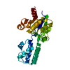
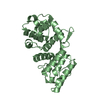
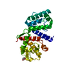
Mass: 92.094 Da / Num. of mol.: 1 / Source method: obtained synthetically / Formula: C3H8O3
#3: Water ChemComp-HOH / water / Water


Mass: 18.015 Da / Num. of mol.: 292 / Source method: isolated from a natural source / Formula: H2O

-
Experimental details

-
Experiment

ExperimentMethod: X-RAY DIFFRACTION / Number of used crystals: 1

-
Sample preparation

CrystalDensity Matthews: 2.41 Å3/Da / Density % sol: 49.03 %
Crystal growTemperature: 295 K / Method: vapor diffusion, sitting drop / pH: 7.2
Details: 50mM Sodium Chloride, 5% Glycerol, 1mM EDTA, 15 - 18% PEG5000-MME
PH range: 7.2 - 7.4

-
Data collection

DiffractionMean temperature: 100 K / Serial crystal experiment: N
Diffraction sourceSource: SYNCHROTRON / Site: APS / Beamline: 17-ID / Wavelength: 1.0719 Å
DetectorType: DECTRIS PILATUS 6M / Detector: PIXEL / Date: Mar 5, 2016 / Details: MIRROR
RadiationMonochromator: Si (III) / Protocol: SINGLE WAVELENGTH / Monochromatic (M) / Laue (L): M / Scattering type: x-ray
Radiation wavelengthWavelength: 1.0719 Å / Relative weight: 1
ReflectionResolution: 1.37→50.83 Å / Num. obs: 52645 / % possible obs: 97.7 % / Redundancy: 5.9 % / CC1/2: 0.99 / Rmerge(I) obs: 0.114 / Rpim(I) all: 0.048 / Net I/σ(I): 17.8
Reflection shellResolution: 1.37→1.39 Å / Redundancy: 3.1 % / Rmerge(I) obs: 0.691 / Mean I/σ(I) obs: 1.44 / Num. unique obs: 2441 / CC1/2: 0.667 / Rpim(I) all: 0.048 / % possible all: 91.9

-
Processing

Software
NameVersionClassification
REFMAC5.8.0238refinement
HKL-2000data reduction
HKL-2000data scaling
PHASERphasing
RefinementMethod to determine structure: MOLECULAR REPLACEMENT
Starting model: 2R0O
Resolution: 1.37→36.7 Å / Cor.coef. Fo:Fc: 0.979 / Cor.coef. Fo:Fc free: 0.971 / SU B: 2.793 / SU ML: 0.047 / Cross valid method: THROUGHOUT / ESU R: 0.052 / ESU R Free: 0.051 / Stereochemistry target values: MAXIMUM LIKELIHOOD / Details: HYDROGENS HAVE BEEN USED IF PRESENT IN THE INPUT
RfactorNum. reflection% reflectionSelection details
Rfree0.1723 2579 4.9 %RANDOM
Rwork0.13416 ---
obs0.13615 49955 97.61 %-
Solvent computationIon probe radii: 0.8 Å / Shrinkage radii: 0.8 Å / VDW probe radii: 1.2 Å / Solvent model: MASK
Displacement parametersBiso mean: 23.031 Å2
Baniso -1Baniso -2Baniso -3
1-3.47 Å20 Å2-0 Å2
2---1.91 Å20 Å2
3----1.57 Å2
Refinement stepCycle: 1 / Resolution: 1.37→36.7 Å
ProteinNucleic acidLigandSolventTotal
Num. atoms1836 0 6 292 2134
Refine LS restraints
Refine-IDTypeDev idealDev ideal targetNumber
X-RAY DIFFRACTIONr_bond_refined_d0.0090.0122000
X-RAY DIFFRACTIONr_bond_other_d0.0030.0171916
X-RAY DIFFRACTIONr_angle_refined_deg1.5341.6352716
X-RAY DIFFRACTIONr_angle_other_deg0.5831.5674459
X-RAY DIFFRACTIONr_dihedral_angle_1_deg5.9365255
X-RAY DIFFRACTIONr_dihedral_angle_2_deg34.92422.589112
X-RAY DIFFRACTIONr_dihedral_angle_3_deg12.16715382
X-RAY DIFFRACTIONr_dihedral_angle_4_deg18.3591514
X-RAY DIFFRACTIONr_chiral_restr0.0870.2262
X-RAY DIFFRACTIONr_gen_planes_refined0.0080.022253
X-RAY DIFFRACTIONr_gen_planes_other0.0010.02425
X-RAY DIFFRACTIONr_nbd_refined
X-RAY DIFFRACTIONr_nbd_other
X-RAY DIFFRACTIONr_nbtor_refined
X-RAY DIFFRACTIONr_nbtor_other
X-RAY DIFFRACTIONr_xyhbond_nbd_refined
X-RAY DIFFRACTIONr_xyhbond_nbd_other
X-RAY DIFFRACTIONr_metal_ion_refined
X-RAY DIFFRACTIONr_metal_ion_other
X-RAY DIFFRACTIONr_symmetry_vdw_refined
X-RAY DIFFRACTIONr_symmetry_vdw_other
X-RAY DIFFRACTIONr_symmetry_hbond_refined
X-RAY DIFFRACTIONr_symmetry_hbond_other
X-RAY DIFFRACTIONr_symmetry_metal_ion_refined
X-RAY DIFFRACTIONr_symmetry_metal_ion_other
X-RAY DIFFRACTIONr_mcbond_it1.7231.911955
X-RAY DIFFRACTIONr_mcbond_other1.7191.907953
X-RAY DIFFRACTIONr_mcangle_it2.182.8791201
X-RAY DIFFRACTIONr_mcangle_other2.1792.8821202
X-RAY DIFFRACTIONr_scbond_it2.8832.3621045
X-RAY DIFFRACTIONr_scbond_other2.8822.3621046
X-RAY DIFFRACTIONr_scangle_it
X-RAY DIFFRACTIONr_scangle_other3.2863.3811505
X-RAY DIFFRACTIONr_long_range_B_refined3.94824.6542402
X-RAY DIFFRACTIONr_long_range_B_other3.75723.6892334
X-RAY DIFFRACTIONr_rigid_bond_restr5.833916
X-RAY DIFFRACTIONr_sphericity_free
X-RAY DIFFRACTIONr_sphericity_bonded
LS refinement shellResolution: 1.371→1.406 Å / Total num. of bins used: 20
RfactorNum. reflection% reflection
Rfree0.348 148 -
Rwork0.294 3432 -
obs--91.65 %

+
About Yorodumi

-
News

-
Feb 9, 2022. New format data for meta-information of EMDB entries

New format data for meta-information of EMDB entries

  • Version 3 of the EMDB header file is now the official format.
  • The previous official version 1.9 will be removed from the archive.

Related info.:EMDB header

External links:wwPDB to switch to version 3 of the EMDB data model

-
Aug 12, 2020. Covid-19 info

Covid-19 info

URL: https://pdbj.org/emnavi/covid19.php

New page: Covid-19 featured information page in EM Navigator.

Related info.:Covid-19 info / Mar 5, 2020. Novel coronavirus structure data

+
Mar 5, 2020. Novel coronavirus structure data

Novel coronavirus structure data

Related info.:Yorodumi Speices / Aug 12, 2020. Covid-19 info

External links:COVID-19 featured content - PDBj / Molecule of the Month (242):Coronavirus Proteases

+
Jan 31, 2019. EMDB accession codes are about to change! (news from PDBe EMDB page)

EMDB accession codes are about to change! (news from PDBe EMDB page)

  • The allocation of 4 digits for EMDB accession codes will soon come to an end. Whilst these codes will remain in use, new EMDB accession codes will include an additional digit and will expand incrementally as the available range of codes is exhausted. The current 4-digit format prefixed with “EMD-” (i.e. EMD-XXXX) will advance to a 5-digit format (i.e. EMD-XXXXX), and so on. It is currently estimated that the 4-digit codes will be depleted around Spring 2019, at which point the 5-digit format will come into force.
  • The EM Navigator/Yorodumi systems omit the EMD- prefix.

Related info.:Q: What is EMD? / ID/Accession-code notation in Yorodumi/EM Navigator

External links:EMDB Accession Codes are Changing Soon! / Contact to PDBj

+
Jul 12, 2017. Major update of PDB

Major update of PDB

  • wwPDB released updated PDB data conforming to the new PDBx/mmCIF dictionary.
  • This is a major update changing the version number from 4 to 5, and with Remediation, in which all the entries are updated.
  • In this update, many items about electron microscopy experimental information are reorganized (e.g. em_software).
  • Now, EM Navigator and Yorodumi are based on the updated data.

External links:wwPDB Remediation / Enriched Model Files Conforming to OneDep Data Standards Now Available in the PDB FTP Archive

-
Yorodumi

Thousand views of thousand structures

  • Yorodumi is a browser for structure data from EMDB, PDB, SASBDB, etc.
  • This page is also the successor to EM Navigator detail page, and also detail information page/front-end page for Omokage search.
  • The word "yorodu" (or yorozu) is an old Japanese word meaning "ten thousand". "mi" (miru) is to see.

Related info.:EMDB / PDB / SASBDB / Comparison of 3 databanks / Yorodumi Search / Aug 31, 2016. New EM Navigator & Yorodumi / Yorodumi Papers / Jmol/JSmol / Function and homology information / Changes in new EM Navigator and Yorodumi

Read more