- PDB-3al3: Crystal Structure of TopBP1 BRCT7/8-BACH1 peptide complex -
+
Open data
ID or keywords:
Loading...
-
Basic information
Entry
Database: PDB / ID: 3al3
Title
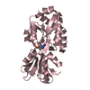
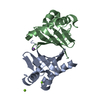
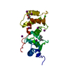
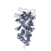
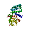
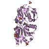
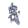
Crystal Structure of TopBP1 BRCT7/8-BACH1 peptide complex
Components
DNA topoisomerase 2-binding protein 1
Peptide of Fanconi anemia group J protein
Keywords
DNA BINDING PROTEIN/PROTEIN BINDING / BRCT domain-phosphopeptide complex / DNA BINDING PROTEIN-PROTEIN BINDING complex
Function / homology
Function and homology information
broken chromosome clustering / BRCA1-B complex / G-quadruplex unwinding activity / Cytosolic iron-sulfur cluster assembly / phosphorylation-dependent protein binding / double-strand break repair involved in meiotic recombination / homologous recombination / DNA replication checkpoint signaling / double-strand break repair via classical nonhomologous end joining / double-strand break repair via alternative nonhomologous end joining ...broken chromosome clustering / BRCA1-B complex / G-quadruplex unwinding activity / Cytosolic iron-sulfur cluster assembly / phosphorylation-dependent protein binding / double-strand break repair involved in meiotic recombination / homologous recombination / DNA replication checkpoint signaling / double-strand break repair via classical nonhomologous end joining / double-strand break repair via alternative nonhomologous end joining / mitotic DNA replication checkpoint signaling / protein-DNA covalent cross-linking repair / protein localization to site of double-strand break / Impaired BRCA2 binding to PALB2 / DNA 5'-3' helicase / chromatin-protein adaptor activity / DNA metabolic process / HDR through Single Strand Annealing (SSA) / response to ionizing radiation / Homologous DNA Pairing and Strand Exchange / Defective homologous recombination repair (HRR) due to BRCA1 loss of function / Defective HDR through Homologous Recombination Repair (HRR) due to PALB2 loss of BRCA1 binding function / Defective HDR through Homologous Recombination Repair (HRR) due to PALB2 loss of BRCA2/RAD51/RAD51C binding function / Resolution of D-loop Structures through Synthesis-Dependent Strand Annealing (SDSA) / mitotic G2 DNA damage checkpoint signaling / Resolution of D-loop Structures through Holliday Junction Intermediates / Impaired BRCA2 binding to RAD51 / chromosome organization / DNA replication initiation / Presynaptic phase of homologous DNA pairing and strand exchange / site of DNA damage / male germ cell nucleus / DNA damage checkpoint signaling / DNA helicase activity / condensed nuclear chromosome / protein serine/threonine kinase activator activity / replication fork / nucleotide-excision repair / PML body / double-strand break repair via homologous recombination / G2/M DNA damage checkpoint / HDR through Homologous Recombination (HRR) / spindle pole / double-strand break repair / chromosome / site of double-strand break / 4 iron, 4 sulfur cluster binding / nuclear membrane / Processing of DNA double-strand break ends / 5'-3' DNA helicase activity / Regulation of TP53 Activity through Phosphorylation / nuclear body / DNA repair / centrosome / regulation of transcription by RNA polymerase II / DNA damage response / ATP hydrolysis activity / DNA binding / nucleoplasm / ATP binding / metal ion binding / identical protein binding / nucleus / cytoplasm Similarity search - Function
Resolution: 2.15→33.9 Å / Cor.coef. Fo:Fc: 0.958 / Cor.coef. Fo:Fc free: 0.939 / WRfactor Rfree: 0.2364 / WRfactor Rwork: 0.1956 / Occupancy max: 1 / Occupancy min: 0.3 / FOM work R set: 0.8292 / SU B: 12.379 / SU ML: 0.146 / SU R Cruickshank DPI: 0.2561 / SU Rfree: 0.1986 / Cross valid method: THROUGHOUT / σ(F): 0 / ESU R Free: 0.199 / Stereochemistry target values: MAXIMUM LIKELIHOOD Details: HYDROGENS HAVE BEEN ADDED IN THE RIDING POSITIONS U VALUES: WITH TLS ADDED
Rfactor
Num. reflection
% reflection
Selection details
Rfree
0.2364
699
5 %
RANDOM
Rwork
0.1955
-
-
-
obs
0.1975
14021
99.86 %
-
Solvent computation
Ion probe radii: 0.8 Å / Shrinkage radii: 0.8 Å / VDW probe radii: 1.4 Å / Solvent model: MASK
In the structure databanks used in Yorodumi, some data are registered as the other names, "COVID-19 virus" and "2019-nCoV". Here are the details of the virus and the list of structure data.
Jan 31, 2019. EMDB accession codes are about to change! (news from PDBe EMDB page)
EMDB accession codes are about to change! (news from PDBe EMDB page)
The allocation of 4 digits for EMDB accession codes will soon come to an end. Whilst these codes will remain in use, new EMDB accession codes will include an additional digit and will expand incrementally as the available range of codes is exhausted. The current 4-digit format prefixed with “EMD-” (i.e. EMD-XXXX) will advance to a 5-digit format (i.e. EMD-XXXXX), and so on. It is currently estimated that the 4-digit codes will be depleted around Spring 2019, at which point the 5-digit format will come into force.
The EM Navigator/Yorodumi systems omit the EMD- prefix.
Related info.:Q: What is EMD? / ID/Accession-code notation in Yorodumi/EM Navigator
Yorodumi is a browser for structure data from EMDB, PDB, SASBDB, etc.
This page is also the successor to EM Navigator detail page, and also detail information page/front-end page for Omokage search.
The word "yorodu" (or yorozu) is an old Japanese word meaning "ten thousand". "mi" (miru) is to see.
Related info.:EMDB / PDB / SASBDB / Comparison of 3 databanks / Yorodumi Search / Aug 31, 2016. New EM Navigator & Yorodumi / Yorodumi Papers / Jmol/JSmol / Function and homology information / Changes in new EM Navigator and Yorodumi
 Movie
Movie Controller


 Open data
 Basic information
 Components
 Keywords
 Function and homology information
 Homo sapiens (human)
 X-RAY DIFFRACTION /
  Authors
 Citation
 Structure visualization
 Downloads & links
 Other downloads
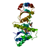


 PDBj
PDBj


 Assembly


 Mass: 46.025 Da / Num. of mol.: 1 / Source method: obtained synthetically / Formula: CH2O2
 Mass: 18.015 Da / Num. of mol.: 96 / Source method: isolated from a natural source / Formula: H2O
 Sample preparation
 / Beamline: 08ID-1
 Processing