[English] 日本語
Yorodumi
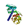
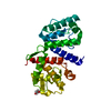
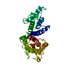
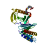
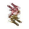
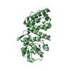
- PDB-1wku: High resolution structure of the human alpha-actinin isoform 3 -

+
Open data


ID or keywords:

Loading...

-
Basic information

Entry
Database: PDB / ID: 1wku
TitleHigh resolution structure of the human alpha-actinin isoform 3
ComponentsAlpha-actinin 3
KeywordsCONTRACTILE PROTEIN / Calponin homology domain / Actin binding domain
Function / homology
Function and homology information


positive regulation of glucose catabolic process to lactate via pyruvate / negative regulation of relaxation of muscle / regulation of the force of skeletal muscle contraction / skeletal muscle atrophy / positive regulation of skeletal muscle fiber development / positive regulation of skeletal muscle tissue growth / response to denervation involved in regulation of muscle adaptation / positive regulation of fast-twitch skeletal muscle fiber contraction / transition between fast and slow fiber / positive regulation of bone mineralization involved in bone maturation ...positive regulation of glucose catabolic process to lactate via pyruvate / negative regulation of relaxation of muscle / regulation of the force of skeletal muscle contraction / skeletal muscle atrophy / positive regulation of skeletal muscle fiber development / positive regulation of skeletal muscle tissue growth / response to denervation involved in regulation of muscle adaptation / positive regulation of fast-twitch skeletal muscle fiber contraction / transition between fast and slow fiber / positive regulation of bone mineralization involved in bone maturation / muscle cell development / negative regulation of oxidative phosphorylation / focal adhesion assembly / Striated Muscle Contraction / bone morphogenesis / negative regulation of glycolytic process / Nephrin family interactions / negative regulation of cold-induced thermogenesis / negative regulation of calcineurin-NFAT signaling cascade / structural constituent of muscle / regulation of aerobic respiration / cortical actin cytoskeleton / pseudopodium / brush border / cell projection / actin filament / Z disc / actin filament binding / integrin binding / cell junction / actin cytoskeleton organization / regulation of apoptotic process / transmembrane transporter binding / focal adhesion / calcium ion binding / extracellular exosome / identical protein binding / plasma membrane / cytosol
Similarity search - Function
Calponin-like domain / EF-hand, Ca insensitive / Ca2+ insensitive EF hand / Ca2+ insensitive EF hand / Actin-binding Protein, T-fimbrin; domain 1 / Spectrin repeat / Spectrin repeat / Spectrin/alpha-actinin / Actinin-type actin-binding domain signature 1. / Actinin-type actin-binding domain signature 2. ...Calponin-like domain / EF-hand, Ca insensitive / Ca2+ insensitive EF hand / Ca2+ insensitive EF hand / Actin-binding Protein, T-fimbrin; domain 1 / Spectrin repeat / Spectrin repeat / Spectrin/alpha-actinin / Actinin-type actin-binding domain signature 1. / Actinin-type actin-binding domain signature 2. / Spectrin repeats / Actinin-type actin-binding domain, conserved site / Calponin homology domain / Calponin homology (CH) domain / Calponin homology domain / CH domain superfamily / Calponin homology (CH) domain profile. / EF-hand, calcium binding motif / EF-hand calcium-binding domain profile. / EF-hand domain / EF-hand domain pair / Orthogonal Bundle / Mainly Alpha
Similarity search - Domain/homology
Biological speciesHomo sapiens (human)
MethodX-RAY DIFFRACTION / SYNCHROTRON / MOLECULAR REPLACEMENT / Resolution: 1.6 Å
AuthorsFranzot, G. / Sjoblom, B. / Gautel, M. / Djinovic Carugo, K.
CitationJournal: J.Mol.Biol. / Year: 2005
Title: The crystal structure of the actin binding domain from alpha-actinin in its closed conformation: structural insight into phospholipid regulation of alpha-actinin
Authors: Franzot, G. / Sjoblom, B. / Gautel, M. / Djinovic Carugo, K.
History
DepositionJun 8, 2004Deposition site: PDBJ / Processing site: PDBJ
Revision 1.0May 17, 2005Provider: repository / Type: Initial release
Revision 1.1Apr 30, 2008Group: Version format compliance
Revision 1.2Jul 13, 2011Group: Version format compliance
Revision 1.3Mar 13, 2024Group: Data collection / Database references
Category: chem_comp_atom / chem_comp_bond ...chem_comp_atom / chem_comp_bond / database_2 / struct_ref_seq_dif
Item: _database_2.pdbx_DOI / _database_2.pdbx_database_accession / _struct_ref_seq_dif.details
Revision 1.4Apr 3, 2024Group: Refinement description / Category: pdbx_initial_refinement_model

-
Structure visualization

Structure viewerMolecule:
MolmilJmol/JSmol

Downloads & links

-
Assembly

Deposited unit
A: Alpha-actinin 3
B: Alpha-actinin 3


Theoretical massNumber of molelcules
Total (without water)58,5512
Polymers58,5512
Non-polymers00
Water8,269459
1
A: Alpha-actinin 3


Theoretical massNumber of molelcules
Total (without water)29,2761
Polymers29,2761
Non-polymers00
Water181
TypeNameSymmetry operationNumber
identity operation1_555x,y,z1
2
B: Alpha-actinin 3


Theoretical massNumber of molelcules
Total (without water)29,2761
Polymers29,2761
Non-polymers00
Water181
TypeNameSymmetry operationNumber
identity operation1_555x,y,z1
Unit cell
Length a, b, c (Å)38.499, 45.960, 66.998
Angle α, β, γ (deg.)86.67, 87.80, 87.33
Int Tables number1
Space group name H-MP1

-
Components

#1: Protein Alpha-actinin 3 / Alpha actinin skeletal muscle isoform 3 / F-actin cross linking protein


Mass: 29275.564 Da / Num. of mol.: 2 / Fragment: actin binding domain (ABD)
Source method: isolated from a genetically manipulated source
Source: (gene. exp.) Homo sapiens (human) / Gene: ACTN3 / Plasmid: pET / Production host: Escherichia coli (E. coli) / Strain (production host): BL21[DE3] / References: UniProt: Q08043
#2: Water ChemComp-HOH / water


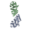
Mass: 18.015 Da / Num. of mol.: 459 / Source method: isolated from a natural source / Formula: H2O

-
Experimental details

-
Experiment

ExperimentMethod: X-RAY DIFFRACTION / Number of used crystals: 1

-
Sample preparation

CrystalDensity Matthews: 2.02 Å3/Da / Density % sol: 39.04 %
Crystal growTemperature: 298 K / Method: vapor diffusion, hanging drop / pH: 5
Details: peg 20000, sodium acetate, pH 5.0, VAPOR DIFFUSION, HANGING DROP, temperature 298.0K

-
Data collection

DiffractionMean temperature: 100 K
Diffraction sourceSource: SYNCHROTRON / Site: ELETTRA / Beamline: 5.2R / Wavelength: 1 Å
DetectorType: MARRESEARCH / Detector: IMAGE PLATE / Details: GRAPHITE MIRRORS
RadiationMonochromator: Si111 / Protocol: SINGLE WAVELENGTH / Monochromatic (M) / Laue (L): M / Scattering type: x-ray
Radiation wavelengthWavelength: 1 Å / Relative weight: 1
ReflectionResolution: 1.6→33.42 Å / Num. all: 56760 / Num. obs: 56760 / % possible obs: 94 % / Observed criterion σ(F): 0 / Observed criterion σ(I): 0 / Biso Wilson estimate: 18.5 Å2
Reflection shellResolution: 1.6→1.7 Å / % possible all: 87.9

-
Processing

Software
NameVersionClassification
CNS1refinement
DENZOdata reduction
SCALEPACKdata scaling
AMoREphasing
RefinementMethod to determine structure: MOLECULAR REPLACEMENT
Starting model: LOW RESOLUTION MODEL OF DIFFERENT SPACE GROUP

Resolution: 1.6→33.42 Å / Rfactor Rfree error: 0.003 / Data cutoff high absF: 197138 / Data cutoff low absF: 0 / Isotropic thermal model: RESTRAINED / Cross valid method: THROUGHOUT / σ(F): 0 / Stereochemistry target values: Engh & Huber
RfactorNum. reflection% reflectionSelection details
Rfree0.208 4024 7.1 %RANDOM
Rwork0.182 ---
obs0.182 56760 94 %-
all-56760 --
Solvent computationSolvent model: FLAT MODEL / Bsol: 46.7633 Å2 / ksol: 0.371786 e/Å3
Displacement parametersBiso mean: 19.8 Å2
Baniso -1Baniso -2Baniso -3
1-3 Å20.73 Å21.39 Å2
2--0.69 Å23.71 Å2
3----3.69 Å2
Refine analyze
FreeObs
Luzzati coordinate error0.19 Å0.16 Å
Luzzati d res low-5 Å
Luzzati sigma a0.09 Å0.07 Å
Refinement stepCycle: LAST / Resolution: 1.6→33.42 Å
ProteinNucleic acidLigandSolventTotal
Num. atoms3626 0 0 459 4085
Refine LS restraints
Refine-IDTypeDev ideal
X-RAY DIFFRACTIONc_bond_d0.004
X-RAY DIFFRACTIONc_bond_d_na
X-RAY DIFFRACTIONc_bond_d_prot
X-RAY DIFFRACTIONc_angle_d
X-RAY DIFFRACTIONc_angle_d_na
X-RAY DIFFRACTIONc_angle_d_prot
X-RAY DIFFRACTIONc_angle_deg1.1
X-RAY DIFFRACTIONc_angle_deg_na
X-RAY DIFFRACTIONc_angle_deg_prot
X-RAY DIFFRACTIONc_dihedral_angle_d19.9
X-RAY DIFFRACTIONc_dihedral_angle_d_na
X-RAY DIFFRACTIONc_dihedral_angle_d_prot
X-RAY DIFFRACTIONc_improper_angle_d0.77
X-RAY DIFFRACTIONc_improper_angle_d_na
X-RAY DIFFRACTIONc_improper_angle_d_prot
X-RAY DIFFRACTIONc_mcbond_it
X-RAY DIFFRACTIONc_mcangle_it
X-RAY DIFFRACTIONc_scbond_it
X-RAY DIFFRACTIONc_scangle_it
LS refinement shellResolution: 1.6→1.7 Å / Rfactor Rfree error: 0.009 / Total num. of bins used: 6
RfactorNum. reflection% reflection
Rfree0.234 626 7.1 %
Rwork0.205 8218 -
obs--87.9 %
Xplor file
Refine-IDSerial noParam fileTopol file
X-RAY DIFFRACTION1PROTEIN_REP.PARAMPROTEIN.TOP
X-RAY DIFFRACTION2WATER_REP.PARAM

+
About Yorodumi

-
News

-
Feb 9, 2022. New format data for meta-information of EMDB entries

New format data for meta-information of EMDB entries

  • Version 3 of the EMDB header file is now the official format.
  • The previous official version 1.9 will be removed from the archive.

Related info.:EMDB header

External links:wwPDB to switch to version 3 of the EMDB data model

-
Aug 12, 2020. Covid-19 info

Covid-19 info

URL: https://pdbjlvh1.pdbj.org/emnavi/covid19.php

New page: Covid-19 featured information page in EM Navigator.

Related info.:Covid-19 info / Mar 5, 2020. Novel coronavirus structure data

+
Mar 5, 2020. Novel coronavirus structure data

Novel coronavirus structure data

Related info.:Yorodumi Speices / Aug 12, 2020. Covid-19 info

External links:COVID-19 featured content - PDBj / Molecule of the Month (242):Coronavirus Proteases

+
Jan 31, 2019. EMDB accession codes are about to change! (news from PDBe EMDB page)

EMDB accession codes are about to change! (news from PDBe EMDB page)

  • The allocation of 4 digits for EMDB accession codes will soon come to an end. Whilst these codes will remain in use, new EMDB accession codes will include an additional digit and will expand incrementally as the available range of codes is exhausted. The current 4-digit format prefixed with “EMD-” (i.e. EMD-XXXX) will advance to a 5-digit format (i.e. EMD-XXXXX), and so on. It is currently estimated that the 4-digit codes will be depleted around Spring 2019, at which point the 5-digit format will come into force.
  • The EM Navigator/Yorodumi systems omit the EMD- prefix.

Related info.:Q: What is EMD? / ID/Accession-code notation in Yorodumi/EM Navigator

External links:EMDB Accession Codes are Changing Soon! / Contact to PDBj

+
Jul 12, 2017. Major update of PDB

Major update of PDB

  • wwPDB released updated PDB data conforming to the new PDBx/mmCIF dictionary.
  • This is a major update changing the version number from 4 to 5, and with Remediation, in which all the entries are updated.
  • In this update, many items about electron microscopy experimental information are reorganized (e.g. em_software).
  • Now, EM Navigator and Yorodumi are based on the updated data.

External links:wwPDB Remediation / Enriched Model Files Conforming to OneDep Data Standards Now Available in the PDB FTP Archive

-
Yorodumi

Thousand views of thousand structures

  • Yorodumi is a browser for structure data from EMDB, PDB, SASBDB, etc.
  • This page is also the successor to EM Navigator detail page, and also detail information page/front-end page for Omokage search.
  • The word "yorodu" (or yorozu) is an old Japanese word meaning "ten thousand". "mi" (miru) is to see.

Related info.:EMDB / PDB / SASBDB / Comparison of 3 databanks / Yorodumi Search / Aug 31, 2016. New EM Navigator & Yorodumi / Yorodumi Papers / Jmol/JSmol / Function and homology information / Changes in new EM Navigator and Yorodumi

Read more