 Movie
Movie Controller


+ Open data

- Basic information
| Entry | Database: PDB / ID: 1dxx | ||||||
|---|---|---|---|---|---|---|---|
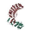
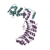
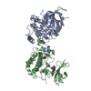
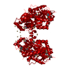
| Title | N-terminal Actin-binding Domain of Human Dystrophin | ||||||
 Components Components | DYSTROPHIN | ||||||
 Keywords Keywords | STRUCTURAL PROTEIN / DYSTROPHIN / MUSCULAR DYSTROPHY / CALPONIN HOMOLOGY DOMAIN / ACTIN-BINDING / UTROPHIN | ||||||
| Function / homology |  Function and homology information Function and homology informationregulation of muscle system process / regulation of cellular response to growth factor stimulus / syntrophin complex / cardiac muscle cell action potential / synaptic signaling / dystrophin-associated glycoprotein complex / cell-substrate junction / motile cilium assembly / peptide biosynthetic process / dystroglycan binding ...regulation of muscle system process / regulation of cellular response to growth factor stimulus / syntrophin complex / cardiac muscle cell action potential / synaptic signaling / dystrophin-associated glycoprotein complex / cell-substrate junction / motile cilium assembly / peptide biosynthetic process / dystroglycan binding / regulation of skeletal muscle contraction by regulation of release of sequestered calcium ion / vinculin binding / regulation of sodium ion transmembrane transport / costamere / Formation of the dystrophin-glycoprotein complex (DGC) / muscle cell development / camera-type eye development / regulation of calcium ion transmembrane transport / Striated Muscle Contraction / filopodium membrane / muscle cell cellular homeostasis / muscle organ development / structural constituent of muscle / myosin binding / maintenance of blood-brain barrier / neuron projection terminus / Non-integrin membrane-ECM interactions / nitric-oxide synthase binding / regulation of skeletal muscle contraction / neuron development / skeletal muscle tissue development / regulation of release of sequestered calcium ion into cytosol by sarcoplasmic reticulum / response to muscle stretch / cardiac muscle contraction / regulation of cardiac muscle contraction by regulation of the release of sequestered calcium ion / positive regulation of neuron differentiation / regulation of heart rate / filopodium / positive regulation of neuron projection development / sarcolemma / structural constituent of cytoskeleton / Z disc / intracellular protein localization / actin binding / protein-containing complex assembly / cytoskeleton / postsynaptic membrane / membrane raft / synapse / cell surface / protein-containing complex / zinc ion binding / nucleus / plasma membrane / cytosol Similarity search - Function | ||||||
| Biological species |  HOMO SAPIENS (human) HOMO SAPIENS (human) | ||||||
| Method |  X-RAY DIFFRACTION / SYNCHROTRON / MOLECULAR REPLACEMENT / Resolution: 2.6 Å X-RAY DIFFRACTION / SYNCHROTRON / MOLECULAR REPLACEMENT / Resolution: 2.6 Å | ||||||
 Authors Authors | Norwood, F.L. / Sutherland-Smith, A.J. / Keep, N.H. / Kendrick-Jones, J. | ||||||
 Citation Citation | Journal: Structure / Year: 2000 Title: The Structure of the N-Terminal Actin-Binding Domain of Human Dystrophin and How Mutations in This Domain May Cause Duchenne or Becker Muscular Dystrophy Authors: Norwood, F.L. / Sutherland-Smith, A.J. / Keep, N.H. / Kendrick-Jones, J. | ||||||
| History |
| ||||||
| Remark 700 | SHEET DETERMINATION METHOD: DSSP THE SHEET IN EACH DIMERIC MOLECULE INVOLVES A STRAND CONTRIBUTED ... SHEET DETERMINATION METHOD: DSSP THE SHEET IN EACH DIMERIC MOLECULE INVOLVES A STRAND CONTRIBUTED FROM EACH COMPONENT CHAIN. |
- Structure visualization
| Structure viewer | Molecule: MolmilJmol/JSmol |
|---|
- Downloads & links
-Download
| PDBx/mmCIF format | 1dxx.cif.gz | 195.2 KB | Display | PDBx/mmCIF format |
|---|---|---|---|---|
| PDB format | pdb1dxx.ent.gz | 156.3 KB | Display | PDB format |
| PDBx/mmJSON format | 1dxx.json.gz | Tree view | PDBx/mmJSON format | |
| Others |  Other downloads Other downloads |
-Validation report
| Arichive directory | https://data.pdbj.org/pub/pdb/validation_reports/dx/1dxxftp://data.pdbj.org/pub/pdb/validation_reports/dx/1dxx | HTTPS FTP |
|---|
-Related structure data
| Related structure data |  1qagS S: Starting model for refinement |
|---|---|
| Similar structure data |


-Links
 PDBj
PDBj


- Assembly
| Deposited unit | 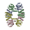
| ||||||||||||||||
|---|---|---|---|---|---|---|---|---|---|---|---|---|---|---|---|---|---|
| 1 | 
| ||||||||||||||||
| 2 | 
| ||||||||||||||||
| Unit cell |
| ||||||||||||||||
| Noncrystallographic symmetry (NCS) | NCS oper:
|
-Components
| #1: Protein | Mass: 28459.275 Da / Num. of mol.: 4 / Fragment: ACTIN-BINDING / Mutation: YES Source method: isolated from a genetically manipulated source Source: (gene. exp.) HOMO SAPIENS (human) / Tissue: MUSCLE / Cellular location: CELL MEMBRANE / Gene: DMD / Plasmid: PGEX-4T2 / Cellular location (production host): CYTOPLASM / Production host:  #2: Water | ChemComp-HOH / |  Mass: 18.015 Da / Num. of mol.: 74 / Source method: isolated from a natural source / Formula: H2O Mass: 18.015 Da / Num. of mol.: 74 / Source method: isolated from a natural source / Formula: H2OSequence details | N-TERMINAL FRAGMENT 1-246 | |
|---|
-Experimental details
-Experiment
| Experiment | Method: X-RAY DIFFRACTION / Number of used crystals: 1 |
|---|
- Sample preparation
| Crystal | Density Matthews: 2.8 Å3/Da / Density % sol: 52 % / Description: DOMAIN SWAPPED SEARCH MODEL USED | ||||||||||||||||||||||||||||||
|---|---|---|---|---|---|---|---|---|---|---|---|---|---|---|---|---|---|---|---|---|---|---|---|---|---|---|---|---|---|---|---|
| Crystal grow | pH: 7.4 / Details: 1.55M AMMONIUM FORMATE, 0.1M HEPES 7.4, pH 7.40 | ||||||||||||||||||||||||||||||
| Crystal | *PLUS Density % sol: 52 % | ||||||||||||||||||||||||||||||
| Crystal grow | *PLUS Temperature: 18 ℃ / Method: vapor diffusion, hanging drop | ||||||||||||||||||||||||||||||
| Components of the solutions | *PLUS
|
-Data collection
| Diffraction | Mean temperature: 100 K |
|---|---|
| Diffraction source | Source: SYNCHROTRON / Site: ELETTRA  / Beamline: 5.2R / Wavelength: 1 / Beamline: 5.2R / Wavelength: 1 |
| Detector | Type: MARRESEARCH / Detector: IMAGE PLATE |
| Radiation | Protocol: SINGLE WAVELENGTH / Monochromatic (M) / Laue (L): M / Scattering type: x-ray |
| Radiation wavelength | Wavelength: 1 Å / Relative weight: 1 |
| Reflection | Resolution: 2.6→40 Å / Num. obs: 36284 / % possible obs: 95.4 % / Redundancy: 1.7 % / Biso Wilson estimate: 52 Å2 / Rmerge(I) obs: 0.051 / Net I/σ(I): 12.7 |
| Reflection shell | Resolution: 2.6→2.69 Å / Redundancy: 1.6 % / Rmerge(I) obs: 0.25 / Mean I/σ(I) obs: 2.6 / % possible all: 91.6 |
| Reflection shell | *PLUS Highest resolution: 2.6 Å / % possible obs: 91.6 % / Num. unique obs: 3448 |
- Processing
| Software |
| ||||||||||||||||||||||||||||||||||||||||||||||||||||||||||||||||||||||||||||||||||||
|---|---|---|---|---|---|---|---|---|---|---|---|---|---|---|---|---|---|---|---|---|---|---|---|---|---|---|---|---|---|---|---|---|---|---|---|---|---|---|---|---|---|---|---|---|---|---|---|---|---|---|---|---|---|---|---|---|---|---|---|---|---|---|---|---|---|---|---|---|---|---|---|---|---|---|---|---|---|---|---|---|---|---|---|---|---|
| Refinement | Method to determine structure: MOLECULAR REPLACEMENT Starting model: PDB ENTRY 1QAG Resolution: 2.6→20 Å / SU B: 5.77 / SU ML: 0.13 / Cross valid method: THROUGHOUT / σ(F): 0 / ESU R: 0.86 / ESU R Free: 0.34 Details: XPLOR BULK SOLVENT MODEL USED TERMINAL RESIDUES FROM PGEX-4T2 CLONING SITE WERE NOT OBSERVED IN MAPS. TIGHT NCS RESTRAINTS WERE IMPOSED DURING ALL STAGES OF REFINEMENT.
| ||||||||||||||||||||||||||||||||||||||||||||||||||||||||||||||||||||||||||||||||||||
| Displacement parameters | Biso mean: 37 Å2
| ||||||||||||||||||||||||||||||||||||||||||||||||||||||||||||||||||||||||||||||||||||
| Refinement step | Cycle: LAST / Resolution: 2.6→20 Å
| ||||||||||||||||||||||||||||||||||||||||||||||||||||||||||||||||||||||||||||||||||||
| Refine LS restraints |
| ||||||||||||||||||||||||||||||||||||||||||||||||||||||||||||||||||||||||||||||||||||
| Software | *PLUS Name: REFMAC / Classification: refinement | ||||||||||||||||||||||||||||||||||||||||||||||||||||||||||||||||||||||||||||||||||||
| Refinement | *PLUS Lowest resolution: 40 Å / Rfactor obs: 0.232 | ||||||||||||||||||||||||||||||||||||||||||||||||||||||||||||||||||||||||||||||||||||
| Solvent computation | *PLUS | ||||||||||||||||||||||||||||||||||||||||||||||||||||||||||||||||||||||||||||||||||||
| Displacement parameters | *PLUS | ||||||||||||||||||||||||||||||||||||||||||||||||||||||||||||||||||||||||||||||||||||
| LS refinement shell | *PLUS Highest resolution: 2.6 Å / Lowest resolution: 2.69 Å / Rfactor Rfree: 0.314 / Rfactor obs: 0.265 |
