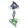
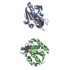
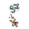
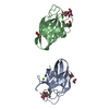
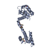
The biological assembly is probably the monomer rather than the dimer. This may be in an open configuration as the chains are in the crystal or in a closed configuration with the regions A31-148 and B153-B256 in the crystal being formed by a single chain
-
Components
#1: Protein
UTROPHINACTINBINDINGREGION
Mass: 26112.334 Da / Num. of mol.: 2 / Fragment: RESIDUES 28-261 / Mutation: SELENOMETHIONINE REPLACES METHIONINE Source method: isolated from a genetically manipulated source Details: The biological assembly is probably the monomer rather than the dimer. This may be in an open configuration as the chains are in the crystal or in a closed configuration with the regions A31- ...Details: The biological assembly is probably the monomer rather than the dimer. This may be in an open configuration as the chains are in the crystal or in a closed configuration with the regions A31-148 and B153-B256 in the crystal being formed by a single chain Source: (gene. exp.) Homo sapiens (human) / Production host: Escherichia coli (E. coli) / References: UniProt: P46939
Resolution: 3→3.16 Å / Redundancy: 2.6 % / Rmerge(I) obs: 0.146 / Mean I/σ(I) obs: 5.1 / Num. unique all: 1772 / % possible all: 97.2
Reflection
*PLUS
Lowest resolution: 24 Å
Reflection shell
*PLUS
% possible obs: 97.2 %
-
Processing
Software
Name
Version
Classification
DENZO
datareduction
SCALEPACK
datascaling
SHELXS
phasing
MLPHARE
phasing
SHARP
phasing
X-PLOR
3.851
refinement
Refinement
Method to determine structure: MAD / Resolution: 3→99 Å / Rfactor Rfree error: 0.01 / Data cutoff high absF: 10000000 / Data cutoff low absF: 0 / Isotropic thermal model: GROUP / Cross valid method: THROUGHOUT / σ(F): 0 / Stereochemistry target values: Engh & Huber Details: Bulk solvent correction and ncs restraints (not strict) used in X-Plor. RESIDUES 31-146 and 156-254 IN CHAINS A AND B USED AS SEPARATE GROUPS WITH WEIGHT 50.0 FOR NCS RESTRAINT.
Rfactor
Num. reflection
% reflection
Selection details
Rfree
0.258
607
5 %
RANDOM
Rwork
0.198
-
-
-
all
-
12253
-
-
obs
-
12253
94.7 %
-
Displacement parameters
Biso mean: 44.5 Å2
Refine analyze
Free
Obs
Luzzati coordinate error
0.42 Å
0.31 Å
Luzzati d res low
-
99 Å
Luzzati sigma a
0.46 Å
0.39 Å
Refinement step
Cycle: LAST / Resolution: 3→99 Å
Protein
Nucleic acid
Ligand
Solvent
Total
Num. atoms
3642
0
0
12
3654
Refine LS restraints
Refine-ID
Type
Dev ideal
X-RAY DIFFRACTION
x_bond_d
0.007
X-RAY DIFFRACTION
x_bond_d_na
X-RAY DIFFRACTION
x_bond_d_prot
X-RAY DIFFRACTION
x_angle_d
X-RAY DIFFRACTION
x_angle_d_na
X-RAY DIFFRACTION
x_angle_d_prot
X-RAY DIFFRACTION
x_angle_deg
1.2
X-RAY DIFFRACTION
x_angle_deg_na
X-RAY DIFFRACTION
x_angle_deg_prot
X-RAY DIFFRACTION
x_dihedral_angle_d
23.1
X-RAY DIFFRACTION
x_dihedral_angle_d_na
X-RAY DIFFRACTION
x_dihedral_angle_d_prot
X-RAY DIFFRACTION
x_improper_angle_d
0.66
X-RAY DIFFRACTION
x_improper_angle_d_na
X-RAY DIFFRACTION
x_improper_angle_d_prot
X-RAY DIFFRACTION
x_mcbond_it
X-RAY DIFFRACTION
x_mcangle_it
X-RAY DIFFRACTION
x_scbond_it
X-RAY DIFFRACTION
x_scangle_it
LS refinement shell
Resolution: 3→3.19 Å / Rfactor Rfree error: 0.034 / Total num. of bins used: 6
In the structure databanks used in Yorodumi, some data are registered as the other names, "COVID-19 virus" and "2019-nCoV". Here are the details of the virus and the list of structure data.
Jan 31, 2019. EMDB accession codes are about to change! (news from PDBe EMDB page)
EMDB accession codes are about to change! (news from PDBe EMDB page)
The allocation of 4 digits for EMDB accession codes will soon come to an end. Whilst these codes will remain in use, new EMDB accession codes will include an additional digit and will expand incrementally as the available range of codes is exhausted. The current 4-digit format prefixed with “EMD-” (i.e. EMD-XXXX) will advance to a 5-digit format (i.e. EMD-XXXXX), and so on. It is currently estimated that the 4-digit codes will be depleted around Spring 2019, at which point the 5-digit format will come into force.
The EM Navigator/Yorodumi systems omit the EMD- prefix.
Related info.:Q: What is EMD? / ID/Accession-code notation in Yorodumi/EM Navigator
Yorodumi is a browser for structure data from EMDB, PDB, SASBDB, etc.
This page is also the successor to EM Navigator detail page, and also detail information page/front-end page for Omokage search.
The word "yorodu" (or yorozu) is an old Japanese word meaning "ten thousand". "mi" (miru) is to see.
Related info.:EMDB / PDB / SASBDB / Comparison of 3 databanks / Yorodumi Search / Aug 31, 2016. New EM Navigator & Yorodumi / Yorodumi Papers / Jmol/JSmol / Function and homology information / Changes in new EM Navigator and Yorodumi
 Movie
Movie Controller


 Open data
 Basic information
 Components
 Keywords
 Function and homology information
 Homo sapiens (human)
 X-RAY DIFFRACTION /
  Authors
 Citation
 Structure visualization
 Downloads & links
 Other downloads
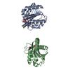

 PDBj
PDBj


 Assembly

 Mass: 18.015 Da / Num. of mol.: 12 / Source method: isolated from a natural source / Formula: H2O
 Sample preparation
 / Beamline: BM14 / Wavelength: 0.9000, 0.9795, 0.9809
 Processing