 Movie
Movie Controller


+ Open data

- Basic information
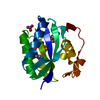
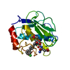
| Entry | Database: PDB / ID: 2oy2 | ||||||
|---|---|---|---|---|---|---|---|
| Title | Human MMP-8 in complex with peptide IAG | ||||||
 Components Components |
| ||||||
 Keywords Keywords | HYDROLASE / MMP-8 / Matrix Metalloproteinase | ||||||
| Function / homology |  Function and homology information Function and homology informationneutrophil collagenase / tumor necrosis factor binding / positive regulation of microglial cell activation / positive regulation of tumor necrosis factor-mediated signaling pathway / positive regulation of neuroinflammatory response / Activation of Matrix Metalloproteinases / endodermal cell differentiation / Collagen degradation / collagen catabolic process / extracellular matrix disassembly ...neutrophil collagenase / tumor necrosis factor binding / positive regulation of microglial cell activation / positive regulation of tumor necrosis factor-mediated signaling pathway / positive regulation of neuroinflammatory response / Activation of Matrix Metalloproteinases / endodermal cell differentiation / Collagen degradation / collagen catabolic process / extracellular matrix disassembly / Degradation of the extracellular matrix / extracellular matrix organization / metalloendopeptidase activity / specific granule lumen / positive regulation of tumor necrosis factor production / tertiary granule lumen / peptidase activity / extracellular matrix / cellular response to lipopolysaccharide / endopeptidase activity / serine-type endopeptidase activity / Neutrophil degranulation / proteolysis / : / extracellular region / zinc ion binding Similarity search - Function | ||||||
| Biological species |  Homo sapiens (human) Homo sapiens (human) | ||||||
| Method |  X-RAY DIFFRACTION / SYNCHROTRON / MOLECULAR REPLACEMENT / Resolution: 1.5 Å X-RAY DIFFRACTION / SYNCHROTRON / MOLECULAR REPLACEMENT / Resolution: 1.5 Å | ||||||
 Authors Authors | Calderone, V. / Bertini, I. / Fragai, M. / Luchinat, C. / Maletta, M. / Yeo, K.J. | ||||||
 Citation Citation | Journal: ANGEW.CHEM.INT.ED.ENGL. / Year: 2006 Title: Snapshots of the reaction mechanism of matrix metalloproteinases. Authors: Bertini, I. / Calderone, V. / Fragai, M. / Luchinat, C. / Maletta, M. / Yeo, K.J. #1: Journal: J.Med.Chem. / Year: 1999Title: Crystal structure of MMP8 complexed with HMR2909 Authors: Matter, H. / Schwab, W. / Barbier, D. / Billen, G. / Haase, B. / Neises, B. / Schudok, M. / Thorwart, W. / Schreuder, H. / Brachvogel, V. | ||||||
| History |
|
- Structure visualization
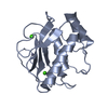
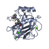
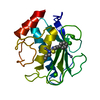
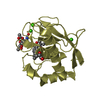
| Structure viewer | Molecule: MolmilJmol/JSmol |
|---|
- Downloads & links
-Download
| PDBx/mmCIF format | 2oy2.cif.gz | 157.8 KB | Display | PDBx/mmCIF format |
|---|---|---|---|---|
| PDB format | pdb2oy2.ent.gz | 122.3 KB | Display | PDB format |
| PDBx/mmJSON format | 2oy2.json.gz | Tree view | PDBx/mmJSON format | |
| Others |  Other downloads Other downloads |
-Validation report
| Arichive directory | https://data.pdbj.org/pub/pdb/validation_reports/oy/2oy2ftp://data.pdbj.org/pub/pdb/validation_reports/oy/2oy2 | HTTPS FTP |
|---|
-Related structure data
| Related structure data |  2oxuC  2oxwC  2oxzC  2oy4C  1bzsS S: Starting model for refinement C: citing same article ( |
|---|---|
| Similar structure data |


-Links
 PDBj
PDBj


- Assembly
| Deposited unit | 
| ||||||||
|---|---|---|---|---|---|---|---|---|---|
| 1 | 
| ||||||||
| 2 | 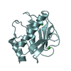
| ||||||||
| Unit cell |
| ||||||||
| Details | The protein is monomeric in vivo but there are two molecules in the asymmetric unit. |
-Components
| #1: Protein | Mass: 17612.115 Da / Num. of mol.: 2 / Fragment: CATALYTIC DOMAIN Source method: isolated from a genetically manipulated source Source: (gene. exp.) Homo sapiens (human) / Gene: MMP8, CLG1 / Plasmid: pET21 / Species (production host): Escherichia coli / Production host:  #2: Protein/peptide | Mass: 259.303 Da / Num. of mol.: 2 / Source method: obtained synthetically #3: Chemical | ChemComp-CA /   Mass: 40.078 Da / Num. of mol.: 4 / Source method: obtained synthetically / Formula: Ca Mass: 40.078 Da / Num. of mol.: 4 / Source method: obtained synthetically / Formula: Ca#4: Chemical | ChemComp-ZN /   Mass: 65.409 Da / Num. of mol.: 4 / Source method: obtained synthetically / Formula: Zn Mass: 65.409 Da / Num. of mol.: 4 / Source method: obtained synthetically / Formula: Zn#5: Water | ChemComp-HOH / |  Mass: 18.015 Da / Num. of mol.: 565 / Source method: isolated from a natural source / Formula: H2O Mass: 18.015 Da / Num. of mol.: 565 / Source method: isolated from a natural source / Formula: H2O |
|---|
-Experimental details
-Experiment
| Experiment | Method: X-RAY DIFFRACTION / Number of used crystals: 1 |
|---|
- Sample preparation
| Crystal | Density Matthews: 2.38 Å3/Da / Density % sol: 48.31 % |
|---|---|
| Crystal grow | Temperature: 298 K / Method: vapor diffusion, sitting drop / pH: 8 Details: 0.1M Tris-HCl, 20% PEG3350, 200mM acetohydroxamic acid, 0.2M MgCl2, pH 8.0, VAPOR DIFFUSION, SITTING DROP, temperature 298K |
-Data collection
| Diffraction | Mean temperature: 100 K |
|---|---|
| Diffraction source | Source: SYNCHROTRON / Site: EMBL/DESY, HAMBURG  / Beamline: BW7A / Wavelength: 0.8423 / Beamline: BW7A / Wavelength: 0.8423 |
| Detector | Type: MARRESEARCH / Detector: CCD / Date: Jul 15, 2006 Details: Fixed exit double crystal Si [111], horizontally focussing |
| Radiation | Monochromator: Fixed exit double crystal Si [111], horizontally focussing Protocol: SINGLE WAVELENGTH / Monochromatic (M) / Laue (L): M / Scattering type: x-ray |
| Radiation wavelength | Wavelength: 0.8423 Å / Relative weight: 1 |
| Reflection | Resolution: 1.5→38.75 Å / Num. all: 55607 / Num. obs: 55607 / % possible obs: 100 % / Observed criterion σ(F): 0 / Observed criterion σ(I): 0 / Redundancy: 6.7 % / Biso Wilson estimate: 10.2 Å2 / Rmerge(I) obs: 0.082 / Rsym value: 0.082 / Net I/σ(I): 6.6 |
| Reflection shell | Resolution: 1.5→1.58 Å / Redundancy: 6 % / Rmerge(I) obs: 0.314 / Mean I/σ(I) obs: 2.2 / Num. unique all: 8108 / Rsym value: 0.314 / % possible all: 100 |
- Processing
| Software |
| ||||||||||||||||||||||||||||||||||||||||||||||||||||||||||||||||||||||||||||||||||||||||||||||||||||||||||||||||||||||||||||||||||||||||||||||||||||||||||||||||||||||||||
|---|---|---|---|---|---|---|---|---|---|---|---|---|---|---|---|---|---|---|---|---|---|---|---|---|---|---|---|---|---|---|---|---|---|---|---|---|---|---|---|---|---|---|---|---|---|---|---|---|---|---|---|---|---|---|---|---|---|---|---|---|---|---|---|---|---|---|---|---|---|---|---|---|---|---|---|---|---|---|---|---|---|---|---|---|---|---|---|---|---|---|---|---|---|---|---|---|---|---|---|---|---|---|---|---|---|---|---|---|---|---|---|---|---|---|---|---|---|---|---|---|---|---|---|---|---|---|---|---|---|---|---|---|---|---|---|---|---|---|---|---|---|---|---|---|---|---|---|---|---|---|---|---|---|---|---|---|---|---|---|---|---|---|---|---|---|---|---|---|---|---|---|
| Refinement | Method to determine structure: MOLECULAR REPLACEMENT Starting model: pdb entry 1BZS Resolution: 1.5→38.75 Å / Cor.coef. Fo:Fc: 0.965 / Cor.coef. Fo:Fc free: 0.955 / SU B: 2.312 / SU ML: 0.041 / Isotropic thermal model: Anisotropic / Cross valid method: THROUGHOUT / σ(F): 0 / σ(I): 0 / ESU R: 0.094 / ESU R Free: 0.074 / Stereochemistry target values: MAXIMUM LIKELIHOOD / Details: HYDROGENS HAVE BEEN ADDED IN THE RIDING POSITIONS
| ||||||||||||||||||||||||||||||||||||||||||||||||||||||||||||||||||||||||||||||||||||||||||||||||||||||||||||||||||||||||||||||||||||||||||||||||||||||||||||||||||||||||||
| Solvent computation | Ion probe radii: 0.8 Å / Shrinkage radii: 0.8 Å / VDW probe radii: 1.4 Å / Solvent model: MASK | ||||||||||||||||||||||||||||||||||||||||||||||||||||||||||||||||||||||||||||||||||||||||||||||||||||||||||||||||||||||||||||||||||||||||||||||||||||||||||||||||||||||||||
| Displacement parameters | Biso mean: 12.388 Å2
| ||||||||||||||||||||||||||||||||||||||||||||||||||||||||||||||||||||||||||||||||||||||||||||||||||||||||||||||||||||||||||||||||||||||||||||||||||||||||||||||||||||||||||
| Refinement step | Cycle: LAST / Resolution: 1.5→38.75 Å
| ||||||||||||||||||||||||||||||||||||||||||||||||||||||||||||||||||||||||||||||||||||||||||||||||||||||||||||||||||||||||||||||||||||||||||||||||||||||||||||||||||||||||||
| Refine LS restraints |
| ||||||||||||||||||||||||||||||||||||||||||||||||||||||||||||||||||||||||||||||||||||||||||||||||||||||||||||||||||||||||||||||||||||||||||||||||||||||||||||||||||||||||||
| LS refinement shell | Resolution: 1.5→1.539 Å / Total num. of bins used: 20
|
