 Movie
Movie Controller

[English] 日本語
 Yorodumi
- PDB-5u94: Crystal structure of the Mycobacterium tuberculosis PASTA kinase ... -
+ Open data

- Basic information
| Entry | Database: PDB / ID: 5u94 | |||||||||
|---|---|---|---|---|---|---|---|---|---|---|
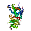
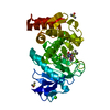
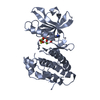
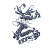
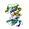
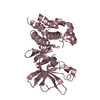
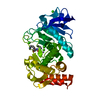
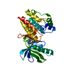
| Title | Crystal structure of the Mycobacterium tuberculosis PASTA kinase PknB in complex with the potential theraputic kinase inhibitor GSK690693. | |||||||||
 Components Components | Serine/threonine-protein kinase PknB | |||||||||
 Keywords Keywords | Transferase/Transferase Inhibitor / Kinase / inhibitor / transferase / Transferase-Transferase Inhibitor complex | |||||||||
| Function / homology |  Function and homology information Function and homology informationnegative regulation of growth rate / negative regulation of fatty acid biosynthetic process / peptidoglycan biosynthetic process / peptidoglycan-based cell wall / manganese ion binding / regulation of cell shape / protein kinase activity / non-specific serine/threonine protein kinase / protein serine kinase activity / protein serine/threonine kinase activity ...negative regulation of growth rate / negative regulation of fatty acid biosynthetic process / peptidoglycan biosynthetic process / peptidoglycan-based cell wall / manganese ion binding / regulation of cell shape / protein kinase activity / non-specific serine/threonine protein kinase / protein serine kinase activity / protein serine/threonine kinase activity / regulation of DNA-templated transcription / ATP binding / identical protein binding / plasma membrane Similarity search - Function | |||||||||
| Biological species |   Mycobacterium tuberculosis (bacteria) Mycobacterium tuberculosis (bacteria) | |||||||||
| Method |  X-RAY DIFFRACTION / SYNCHROTRON / MOLECULAR REPLACEMENT / Resolution: 2.2 Å X-RAY DIFFRACTION / SYNCHROTRON / MOLECULAR REPLACEMENT / Resolution: 2.2 Å | |||||||||
 Authors Authors | Wlodarchak, N. / Satyshur, K. / Striker, R. | |||||||||
| Funding support |  United States, 2items United States, 2items
| |||||||||
 Citation Citation | Journal: Mol. Pharm. / Year: 2018 Title: In Silico Screen and Structural Analysis Identifies Bacterial Kinase Inhibitors which Act with beta-Lactams To Inhibit Mycobacterial Growth. Authors: Wlodarchak, N. / Teachout, N. / Beczkiewicz, J. / Procknow, R. / Schaenzer, A.J. / Satyshur, K. / Pavelka, M. / Zuercher, W. / Drewry, D. / Sauer, J.D. / Striker, R. | |||||||||
| History |
|
- Structure visualization
| Structure viewer | Molecule: MolmilJmol/JSmol |
|---|
- Downloads & links
-Download
| PDBx/mmCIF format | 5u94.cif.gz | 111.1 KB | Display | PDBx/mmCIF format |
|---|---|---|---|---|
| PDB format | pdb5u94.ent.gz | 85.1 KB | Display | PDB format |
| PDBx/mmJSON format | 5u94.json.gz | Tree view | PDBx/mmJSON format | |
| Others |  Other downloads Other downloads |
-Validation report
| Arichive directory | https://data.pdbj.org/pub/pdb/validation_reports/u9/5u94ftp://data.pdbj.org/pub/pdb/validation_reports/u9/5u94 | HTTPS FTP |
|---|
-Related structure data
| Related structure data | 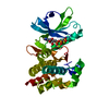 1o6yS S: Starting model for refinement |
|---|---|
| Similar structure data |


-Links
 PDBj
PDBj- Assembly
| Deposited unit | 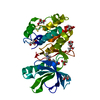
| ||||||||
|---|---|---|---|---|---|---|---|---|---|
| 1 |
| ||||||||
| Unit cell |
| ||||||||
| Components on special symmetry positions |
|
-Components
| #1: Protein | Mass: 30827.719 Da / Num. of mol.: 1 / Fragment: UNP residues 1-280 Source method: isolated from a genetically manipulated source Source: (gene. exp.) Mycobacterium tuberculosis (strain ATCC 25618 / H37Rv) (bacteria)Strain: ATCC 25618 / H37Rv / Gene: pknB, Rv0014c, MTCY10H4.14c / Production host: References: UniProt: P9WI81, non-specific serine/threonine protein kinase | ||||
|---|---|---|---|---|---|
| #2: Chemical | ChemComp-G93 /   Mass: 425.484 Da / Num. of mol.: 1 / Source method: obtained synthetically / Formula: C21H27N7O3 Mass: 425.484 Da / Num. of mol.: 1 / Source method: obtained synthetically / Formula: C21H27N7O3 | ||||
| #3: Chemical | ChemComp-GOL /   Mass: 92.094 Da / Num. of mol.: 8 / Source method: obtained synthetically / Formula: C3H8O3 Mass: 92.094 Da / Num. of mol.: 8 / Source method: obtained synthetically / Formula: C3H8O3#4: Chemical | ChemComp-MG / |   Mass: 24.305 Da / Num. of mol.: 1 / Source method: obtained synthetically / Formula: Mg Mass: 24.305 Da / Num. of mol.: 1 / Source method: obtained synthetically / Formula: Mg#5: Water | ChemComp-HOH / |  Mass: 18.015 Da / Num. of mol.: 44 / Source method: isolated from a natural source / Formula: H2O Mass: 18.015 Da / Num. of mol.: 44 / Source method: isolated from a natural source / Formula: H2O |
-Experimental details
-Experiment
| Experiment | Method: X-RAY DIFFRACTION / Number of used crystals: 1 |
|---|
- Sample preparation
| Crystal | Density Matthews: 2.8 Å3/Da / Density % sol: 56.01 % Description: Irregular rectangle or arrowhead shape, 0.05-0.1mm average size. |
|---|---|
| Crystal grow | Temperature: 296 K / Method: vapor diffusion, sitting drop / pH: 7.5 Details: 12.5% PEG 3350, 0.25M bis-tris propane pH 7.5, 0.5% glycerol, 0.1% thymidne, and 0.1% b-cyclodextran all added 1:1 with protein (9.8mg/mL) and 120uM inhibitor in 150mM NaCl, 10mM Tris pH 8. ...Details: 12.5% PEG 3350, 0.25M bis-tris propane pH 7.5, 0.5% glycerol, 0.1% thymidne, and 0.1% b-cyclodextran all added 1:1 with protein (9.8mg/mL) and 120uM inhibitor in 150mM NaCl, 10mM Tris pH 8.0, 1mM DTT, ans 1mM MgCl2 |
-Data collection
| Diffraction | Mean temperature: 100 K |
|---|---|
| Diffraction source | Source: SYNCHROTRON / Site: APS / Beamline: 21-ID-D / Wavelength: 1.12717 Å |
| Detector | Type: DECTRIS EIGER X 9M / Detector: PIXEL / Date: Aug 23, 2015 |
| Radiation | Monochromator: Si(111) / Protocol: SINGLE WAVELENGTH / Monochromatic (M) / Laue (L): M / Scattering type: x-ray |
| Radiation wavelength | Wavelength: 1.12717 Å / Relative weight: 1 |
| Reflection | Resolution: 2.05→50 Å / Num. obs: 20754 / % possible obs: 96.5 % / Redundancy: 12.7 % / Rsym value: 0.105 / Net I/σ(I): 23.59 |
| Reflection shell | Resolution: 2.05→2.09 Å / Num. unique all: 748 / Rpim(I) all: 0.048 / % possible all: 77.7 |
- Processing
| Software |
| |||||||||||||||||||||||||||||||||||||||||||||||||||||||||||||||||||||||||||||||||||||||||||
|---|---|---|---|---|---|---|---|---|---|---|---|---|---|---|---|---|---|---|---|---|---|---|---|---|---|---|---|---|---|---|---|---|---|---|---|---|---|---|---|---|---|---|---|---|---|---|---|---|---|---|---|---|---|---|---|---|---|---|---|---|---|---|---|---|---|---|---|---|---|---|---|---|---|---|---|---|---|---|---|---|---|---|---|---|---|---|---|---|---|---|---|---|
| Refinement | Method to determine structure: MOLECULAR REPLACEMENT Starting model: 1O6Y Resolution: 2.2→41.686 Å / SU ML: 0.22 / Cross valid method: FREE R-VALUE / σ(F): 1.34 / Phase error: 33.04
| |||||||||||||||||||||||||||||||||||||||||||||||||||||||||||||||||||||||||||||||||||||||||||
| Solvent computation | Shrinkage radii: 0.9 Å / VDW probe radii: 1.11 Å | |||||||||||||||||||||||||||||||||||||||||||||||||||||||||||||||||||||||||||||||||||||||||||
| Displacement parameters | Biso max: 238.26 Å2 / Biso mean: 83.8267 Å2 / Biso min: 45.71 Å2 | |||||||||||||||||||||||||||||||||||||||||||||||||||||||||||||||||||||||||||||||||||||||||||
| Refinement step | Cycle: final / Resolution: 2.2→41.686 Å
| |||||||||||||||||||||||||||||||||||||||||||||||||||||||||||||||||||||||||||||||||||||||||||
| Refine LS restraints |
| |||||||||||||||||||||||||||||||||||||||||||||||||||||||||||||||||||||||||||||||||||||||||||
| LS refinement shell | Refine-ID: X-RAY DIFFRACTION / Total num. of bins used: 12
|
