Movie
Movie Controller

[English] 日本語
 Yorodumi
- PDB-6n7w: Structure of bacteriophage T7 leading-strand DNA polymerase (D5A/... -
+ Open data

- Basic information
| Entry | Database: PDB / ID: 6n7w | ||||||
|---|---|---|---|---|---|---|---|
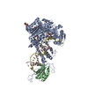
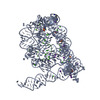
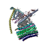
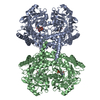
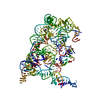
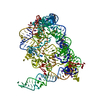
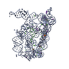
| Title | Structure of bacteriophage T7 leading-strand DNA polymerase (D5A/E7A)/Trx in complex with a DNA fork and incoming dTTP (from multiple lead complexes) | ||||||
 Components Components |
| ||||||
 Keywords Keywords | TRANSFERASE/DNA / DNA polymerase / helicase / DNA replication / replisome / TRANSFERASE-DNA complex | ||||||
| Function / homology |  Function and homology information Function and homology informationDNA synthesis involved in DNA replication / DNA exonuclease activity / viral DNA genome replication / Hydrolases; Acting on ester bonds; Exodeoxyribonucleases producing 5'-phosphomonoesters / DNA polymerase processivity factor activity / protein-disulfide reductase activity / 3'-5' exonuclease activity / cell redox homeostasis / DNA-templated DNA replication / double-strand break repair ...DNA synthesis involved in DNA replication / DNA exonuclease activity / viral DNA genome replication / Hydrolases; Acting on ester bonds; Exodeoxyribonucleases producing 5'-phosphomonoesters / DNA polymerase processivity factor activity / protein-disulfide reductase activity / 3'-5' exonuclease activity / cell redox homeostasis / DNA-templated DNA replication / double-strand break repair / DNA-directed DNA polymerase / DNA-directed DNA polymerase activity / nucleotide binding / DNA binding / metal ion binding / cytosol / cytoplasm Similarity search - Function | ||||||
| Biological species |   Enterobacteria phage T7 (virus) Enterobacteria phage T7 (virus) | ||||||
| Method | ELECTRON MICROSCOPY / single particle reconstruction / cryo EM / Resolution: 4.5 Å | ||||||
 Authors Authors | Gao, Y. / Fox, T. / Val, N. / Yang, W. | ||||||
| Funding support |  United States, 1items United States, 1items
| ||||||
 Citation Citation | Journal: Science / Year: 2019 Title: Structures and operating principles of the replisome. Authors: Yang Gao / Yanxiang Cui / Tara Fox / Shiqiang Lin / Huaibin Wang / Natalia de Val / Z Hong Zhou / Wei Yang / Abstract: Visualization in atomic detail of the replisome that performs concerted leading- and lagging-DNA strand synthesis at a replication fork has not been reported. Using bacteriophage T7 as a model ...Visualization in atomic detail of the replisome that performs concerted leading- and lagging-DNA strand synthesis at a replication fork has not been reported. Using bacteriophage T7 as a model system, we determined cryo-electron microscopy structures up to 3.2-angstroms resolution of helicase translocating along DNA and of helicase-polymerase-primase complexes engaging in synthesis of both DNA strands. Each domain of the spiral-shaped hexameric helicase translocates sequentially hand-over-hand along a single-stranded DNA coil, akin to the way AAA+ ATPases (adenosine triphosphatases) unfold peptides. Two lagging-strand polymerases are attached to the primase, ready for Okazaki fragment synthesis in tandem. A β hairpin from the leading-strand polymerase separates two parental DNA strands into a T-shaped fork, thus enabling the closely coupled helicase to advance perpendicular to the downstream DNA duplex. These structures reveal the molecular organization and operating principles of a replisome. | ||||||
| History |
|
- Structure visualization
| Movie |
Movie viewer |
|---|---|
| Structure viewer | Molecule: MolmilJmol/JSmol |
- Downloads & links
-Download
| PDBx/mmCIF format | 6n7w.cif.gz | 191.9 KB | Display | PDBx/mmCIF format |
|---|---|---|---|---|
| PDB format | pdb6n7w.ent.gz | 144.7 KB | Display | PDB format |
| PDBx/mmJSON format | 6n7w.json.gz | Tree view | PDBx/mmJSON format | |
| Others |  Other downloads Other downloads |
-Validation report
| Arichive directory | https://data.pdbj.org/pub/pdb/validation_reports/n7/6n7wftp://data.pdbj.org/pub/pdb/validation_reports/n7/6n7w | HTTPS FTP |
|---|
-Related structure data
| Related structure data | 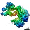 0365MC 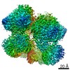 0357C  0359C 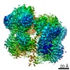 0362C 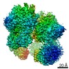 0363C 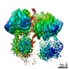 0364C  0379C 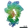 0380C 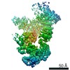 0381C  0382C 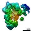 0386C 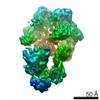 0387C 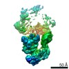 0388C 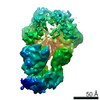 0389C 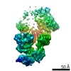 0390C 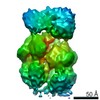 0391C 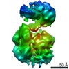 0392C 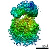 0393C 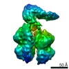 0394C 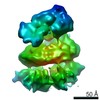 0395C  6n7iC  6n7nC 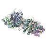 6n7sC 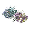 6n7tC 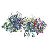 6n7vC 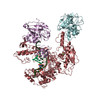 6n9uC 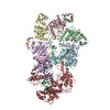 6n9vC 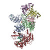 6n9wC  6n9xC M: map data used to model this data C: citing same article ( |
|---|---|
| Similar structure data |

-Links
 PDBj
PDBj


- Assembly
| Deposited unit | 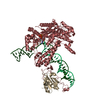
|
|---|---|
| 1 |
|
-Components
-Protein , 2 types, 2 molecules HI
| #1: Protein | Mass: 79805.625 Da / Num. of mol.: 1 / Mutation: D5A, E7A Source method: isolated from a genetically manipulated source Source: (gene. exp.) Enterobacteria phage T7 (virus) / Production host: References: UniProt: P00581, DNA-directed DNA polymerase, Hydrolases; Acting on ester bonds; Exodeoxyribonucleases producing 5'-phosphomonoesters |
|---|---|
| #2: Protein | Mass: 11818.582 Da / Num. of mol.: 1 Source method: isolated from a genetically manipulated source Source: (gene. exp.) |
-DNA chain , 2 types, 2 molecules PT
| #3: DNA chain | Mass: 7716.011 Da / Num. of mol.: 1 / Source method: obtained synthetically / Source: (synth.) Enterobacteria phage T7 (virus) |
|---|---|
| #4: DNA chain | Mass: 23278.844 Da / Num. of mol.: 1 / Source method: obtained synthetically / Source: (synth.) Enterobacteria phage T7 (virus) |
-Non-polymers , 2 types, 2 molecules 

| #5: Chemical | ChemComp-MG /  Mass: 24.305 Da / Num. of mol.: 1 / Source method: obtained synthetically / Formula: Mg Mass: 24.305 Da / Num. of mol.: 1 / Source method: obtained synthetically / Formula: Mg |
|---|---|
| #6: Chemical | ChemComp-TTP /  Mass: 482.168 Da / Num. of mol.: 1 / Source method: obtained synthetically / Formula: C10H17N2O14P3 Mass: 482.168 Da / Num. of mol.: 1 / Source method: obtained synthetically / Formula: C10H17N2O14P3 |
-Experimental details
-Experiment
| Experiment | Method: ELECTRON MICROSCOPY |
|---|---|
| EM experiment | Aggregation state: PARTICLE / 3D reconstruction method: single particle reconstruction |
- Sample preparation
| Component | Name: D5AE7A mutant gp5 DNA polymerase complexed with trx, fork DNA, and incoming dTTP (from gp4-gp5 leading-strand complexes) Type: COMPLEX / Entity ID: #1-#4 / Source: RECOMBINANT | ||||||||||||||||||||
|---|---|---|---|---|---|---|---|---|---|---|---|---|---|---|---|---|---|---|---|---|---|
| Source (natural) | Organism: Enterobacteria phage T7 (virus) | ||||||||||||||||||||
| Source (recombinant) | Organism: | ||||||||||||||||||||
| Buffer solution | pH: 7.5 | ||||||||||||||||||||
| Buffer component |
| ||||||||||||||||||||
| Specimen | Embedding applied: NO / Shadowing applied: NO / Staining applied: NO / Vitrification applied: YES | ||||||||||||||||||||
| Specimen support | Details: unspecified | ||||||||||||||||||||
| Vitrification | Instrument: FEI VITROBOT MARK I / Cryogen name: ETHANE / Humidity: 100 % / Chamber temperature: 295 K |
- Electron microscopy imaging
| Experimental equipment |  Model: Titan Krios / Image courtesy: FEI Company |
|---|---|
| Microscopy | Model: FEI TITAN KRIOS |
| Electron gun | Electron source:  FIELD EMISSION GUN / Accelerating voltage: 300 kV / Illumination mode: FLOOD BEAM FIELD EMISSION GUN / Accelerating voltage: 300 kV / Illumination mode: FLOOD BEAM |
| Electron lens | Mode: BRIGHT FIELD |
| Image recording | Electron dose: 40 e/Å2 / Detector mode: COUNTING / Film or detector model: GATAN K2 SUMMIT (4k x 4k) |
- Processing
| EM software |
| ||||||||||||||||||
|---|---|---|---|---|---|---|---|---|---|---|---|---|---|---|---|---|---|---|---|
| CTF correction | Type: PHASE FLIPPING AND AMPLITUDE CORRECTION | ||||||||||||||||||
| 3D reconstruction | Resolution: 4.5 Å / Resolution method: FSC 0.143 CUT-OFF / Num. of particles: 129003 / Symmetry type: POINT | ||||||||||||||||||
| Atomic model building | Protocol: FLEXIBLE FIT / Space: REAL | ||||||||||||||||||
| Atomic model building | PDB-ID: 1T8E Accession code: 1T8E / Source name: PDB / Type: experimental model |