 Movie
Movie Controller


+ Open data

- Basic information
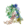
| Entry | Database: PDB / ID: 3bwp | ||||||
|---|---|---|---|---|---|---|---|
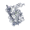
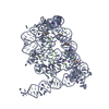
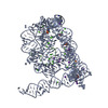
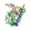
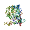
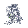
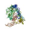
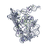
| Title | Crystal structure of a self-spliced group II intron | ||||||
 Components Components | Group IIC intron | ||||||
 Keywords Keywords | RNA / Ribonucleic acid / Intron / Group II | ||||||
| Function / homology | : / : / RNA / RNA (> 10) / RNA (> 100) Function and homology information Function and homology information | ||||||
| Method |  X-RAY DIFFRACTION / SYNCHROTRON / MAD / Resolution: 3.1 Å X-RAY DIFFRACTION / SYNCHROTRON / MAD / Resolution: 3.1 Å | ||||||
 Authors Authors | Toor, N. / Keating, K.S. / Taylor, S.D. / Pyle, A.M. | ||||||
 Citation Citation | Journal: Science / Year: 2008 Title: Crystal structure of a self-spliced group II intron Authors: Toor, N. / Keating, K.S. / Taylor, S.D. / Pyle, A.M. | ||||||
| History |
|
- Structure visualization
| Structure viewer | Molecule: MolmilJmol/JSmol |
|---|
- Downloads & links
-Download
| PDBx/mmCIF format | 3bwp.cif.gz | 202.9 KB | Display | PDBx/mmCIF format |
|---|---|---|---|---|
| PDB format | pdb3bwp.ent.gz | 153.4 KB | Display | PDB format |
| PDBx/mmJSON format | 3bwp.json.gz | Tree view | PDBx/mmJSON format | |
| Others |  Other downloads Other downloads |
-Validation report
| Arichive directory | https://data.pdbj.org/pub/pdb/validation_reports/bw/3bwpftp://data.pdbj.org/pub/pdb/validation_reports/bw/3bwp | HTTPS FTP |
|---|
-Related structure data
| Similar structure data |
|---|

-Links
 PDBj
PDBj


- Assembly
| Deposited unit | 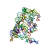
| ||||||||
|---|---|---|---|---|---|---|---|---|---|
| 1 |
| ||||||||
| Unit cell |
|
-Components
| #1: RNA chain | Mass: 133652.156 Da / Num. of mol.: 1 / Source method: obtained synthetically Details: sequence occurs naturally in Oceanobacillus iheyensis. Sequence modified using PCR References: GenBank: 42632302 | ||||
|---|---|---|---|---|---|
| #2: Chemical | ChemComp-MG /   Mass: 24.305 Da / Num. of mol.: 7 / Source method: obtained synthetically / Formula: Mg Mass: 24.305 Da / Num. of mol.: 7 / Source method: obtained synthetically / Formula: Mg#3: Chemical |   Mass: 39.098 Da / Num. of mol.: 2 / Source method: obtained synthetically / Formula: K Mass: 39.098 Da / Num. of mol.: 2 / Source method: obtained synthetically / Formula: K#4: Water | ChemComp-HOH / |  Mass: 18.015 Da / Num. of mol.: 8 / Source method: isolated from a natural source / Formula: H2O Mass: 18.015 Da / Num. of mol.: 8 / Source method: isolated from a natural source / Formula: H2O |
-Experimental details
-Experiment
| Experiment | Method: X-RAY DIFFRACTION / Number of used crystals: 2 |
|---|
- Sample preparation
| Crystal | Density Matthews: 3.57 Å3/Da / Density % sol: 65.5 % | ||||||||||||||||||||||||||||||||||||
|---|---|---|---|---|---|---|---|---|---|---|---|---|---|---|---|---|---|---|---|---|---|---|---|---|---|---|---|---|---|---|---|---|---|---|---|---|---|
| Crystal grow | Temperature: 303 K / Method: vapor diffusion, sitting drop / pH: 7 Details: 6% PEG 3350, 150 mM magnesium acetate, 350 mM KCl, 50 mM Na-HEPES pH 7.0, 0.5 mM spermine, VAPOR DIFFUSION, SITTING DROP, temperature 303K | ||||||||||||||||||||||||||||||||||||
| Components of the solutions |
|
-Data collection
| Diffraction | Mean temperature: 100 K |
|---|---|
| Diffraction source | Source: SYNCHROTRON / Site: APS  / Beamline: 24-ID-C / Wavelength: 0.9795 Å / Beamline: 24-ID-C / Wavelength: 0.9795 Å |
| Detector | Type: ADSC QUANTUM 315 / Detector: CCD / Date: Jun 17, 2007 |
| Radiation | Monochromator: Silicon / Protocol: SINGLE WAVELENGTH / Monochromatic (M) / Laue (L): M / Scattering type: x-ray |
| Radiation wavelength | Wavelength: 0.9795 Å / Relative weight: 1 |
| Reflection | Resolution: 3.1→50 Å / Num. obs: 34541 / % possible obs: 99.6 % / Observed criterion σ(F): 0 / Observed criterion σ(I): 0 / Redundancy: 14.8 % / Rmerge(I) obs: 0.149 / Χ2: 0.988 / Net I/σ(I): 12.3 |
| Reflection shell | Resolution: 3.1→3.21 Å / Redundancy: 6.2 % / Rmerge(I) obs: 0.439 / Mean I/σ(I) obs: 3.7 / Num. unique all: 3365 / Χ2: 1.023 / % possible all: 98.7 |
-Phasing
| Phasing | Method: MAD |
|---|
- Processing
| Software |
| ||||||||||||||||||||||||||||||||||||||||||||
|---|---|---|---|---|---|---|---|---|---|---|---|---|---|---|---|---|---|---|---|---|---|---|---|---|---|---|---|---|---|---|---|---|---|---|---|---|---|---|---|---|---|---|---|---|---|
| Refinement | Method to determine structure: MAD / Resolution: 3.1→50 Å / Isotropic thermal model: isotropic / Cross valid method: THROUGHOUT / σ(F): 0 / σ(I): 0 Stereochemistry target values: Parkinson, Vojtechovsky, Clowney, Brunger & Berman
| ||||||||||||||||||||||||||||||||||||||||||||
| Displacement parameters | Biso mean: 79.413 Å2 | ||||||||||||||||||||||||||||||||||||||||||||
| Refine analyze |
| ||||||||||||||||||||||||||||||||||||||||||||
| Refinement step | Cycle: LAST / Resolution: 3.1→50 Å
| ||||||||||||||||||||||||||||||||||||||||||||
| Refine LS restraints |
| ||||||||||||||||||||||||||||||||||||||||||||
| LS refinement shell | Resolution: 3.1→3.21 Å
|
