Yang Gao / Yanxiang Cui / Tara Fox / Shiqiang Lin / Huaibin Wang / Natalia de Val / Z Hong Zhou / Wei Yang /
PubMed Abstract
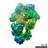
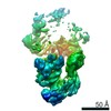
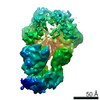
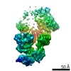
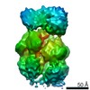
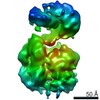
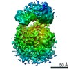
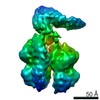
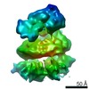
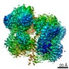
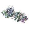
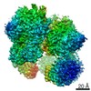
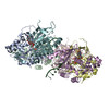
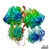
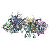
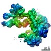
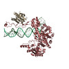
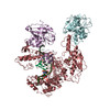
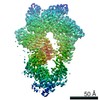
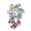
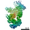
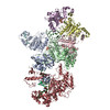
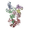
Visualization in atomic detail of the replisome that performs concerted leading- and lagging-DNA strand synthesis at a replication fork has not been reported. Using bacteriophage T7 as a model ...Visualization in atomic detail of the replisome that performs concerted leading- and lagging-DNA strand synthesis at a replication fork has not been reported. Using bacteriophage T7 as a model system, we determined cryo-electron microscopy structures up to 3.2-angstroms resolution of helicase translocating along DNA and of helicase-polymerase-primase complexes engaging in synthesis of both DNA strands. Each domain of the spiral-shaped hexameric helicase translocates sequentially hand-over-hand along a single-stranded DNA coil, akin to the way AAA+ ATPases (adenosine triphosphatases) unfold peptides. Two lagging-strand polymerases are attached to the primase, ready for Okazaki fragment synthesis in tandem. A β hairpin from the leading-strand polymerase separates two parental DNA strands into a T-shaped fork, thus enabling the closely coupled helicase to advance perpendicular to the downstream DNA duplex. These structures reveal the molecular organization and operating principles of a replisome.
EMDB-0357, PDB-6n7i: Structure of bacteriophage T7 E343Q mutant gp4 helicase-primase in complex with ssDNA, dTTP, AC dinucleotide and CTP (gp4(5)-DNA) Method: EM (single particle) / Resolution: 3.2 Å
EMDB-0359, PDB-6n7n: Structure of bacteriophage T7 E343Q mutant gp4 helicase-primase in complex with ssDNA, dTTP, AC dinucleotide and CTP (form I) Method: EM (single particle) / Resolution: 3.5 Å
EMDB-0362, PDB-6n7s: Structure of bacteriophage T7 E343Q mutant gp4 helicase-primase in complex with ssDNA, dTTP, AC dinucleotide and CTP (form II) Method: EM (single particle) / Resolution: 4.6 Å
EMDB-0363, PDB-6n7t: Structure of bacteriophage T7 E343Q mutant gp4 helicase-primase in complex with ssDNA, dTTP, AC dinucleotide and CTP (form III) Method: EM (single particle) / Resolution: 3.9 Å
EMDB-0364, PDB-6n7v: Structure of bacteriophage T7 gp4 (helicase-primase, E343Q mutant) in complex with ssDNA, dTTP, AC dinucleotide, and CTP (from multiple lead complexes) Method: EM (single particle) / Resolution: 3.8 Å
EMDB-0365, PDB-6n7w: Structure of bacteriophage T7 leading-strand DNA polymerase (D5A/E7A)/Trx in complex with a DNA fork and incoming dTTP (from multiple lead complexes) Method: EM (single particle) / Resolution: 4.5 Å
EMDB-0379, PDB-6n9u: Structure of bacteriophage T7 lagging-strand DNA polymerase (D5A/E7A) interacting with primase domains of two gp4 subunits bound to an RNA/DNA hybrid and dTTP (from LagS1) Method: EM (single particle) / Resolution: 3.7 Å
EMDB-0380, PDB-6n9v: Structure of bacteriophage T7 lagging-strand DNA polymerase (D5A/E7A) and gp4 (helicase/primase) bound to DNA including RNA/DNA hybrid, and an incoming dTTP (LagS1) Method: EM (single particle) / Resolution: 4.0 Å
EMDB-0381, PDB-6n9w: Structure of bacteriophage T7 lagging-strand DNA polymerase (D5A/E7A) and gp4 (helicase/primase) bound to DNA including RNA/DNA hybrid, and an incoming dTTP (LagS2) Method: EM (single particle) / Resolution: 4.0 Å
EMDB-0382, PDB-6n9x: Structure of bacteriophage T7 lagging-strand DNA polymerase (D5A/E7A) and gp4 (helicase/primase) bound to DNA including RNA/DNA hybrid, and an incoming dTTP (LagS3) Method: EM (single particle) / Resolution: 4.1 Å
EMDB-0386: Structure of two bacteriophage T7 lagging-strand DNA polymerase (D5A/E7A )/Trx interacting with primase domains, one Pol with RNA/DNA hybrid, and dTTP interacting and the second Pol in apo form (LagS4) Method: EM (single particle) / Resolution: 8.6 Å
EMDB-0387: Structure of bacteriophage T7 lagging-strand DNA polymerase (D5A/E7A)/Trx interacting with primase domains of two gp4 subunits (E and F), with gp4 helicase bound to a DNA fork and dTTP (LagL1) Method: EM (single particle) / Resolution: 6.6 Å
EMDB-0388: Structure of bacteriophage T7 lagging-strand DNA polymerase (D5A/E7A)/Trx interacting with primase domains of two gp4 subunits (C and D), with gp4 helicase bound to a DNA fork and dTTP (LagL2) Method: EM (single particle) / Resolution: 7.1 Å
EMDB-0389: Structure of bacteriophage T7 lagging-strand DNA polymerase (D5A/E7A)/Trx interacting with primase domains of two gp4 subunits (B and C), with gp4 helicase bound to a DNA fork and dTTP (LagL3) Method: EM (single particle) / Resolution: 7.8 Å
EMDB-0390: Structure of bacteriophage T7 lagging-strand DNA polymerase (D5A/E7A)/Trx interacting with primase domains of two gp4 subunits (A and B), with gp4 helicase bound to a DNA fork and dTTP (LagL4) Method: EM (single particle) / Resolution: 8.3 Å
EMDB-0391: Structure of bacteriophage T7 leading-strand DNA polymerase (D5A/E7A)/Trx and of gp4 (E343Q) bound to a DNA fork (Lead1) Method: EM (single particle) / Resolution: 11.2 Å
EMDB-0392: Structure of bacteriophage T7 leading-strand DNA polymerase (D5A/E7A)/Trx and of gp4 (E343Q) bound to a DNA fork (Lead2) Method: EM (single particle) / Resolution: 13.3 Å
EMDB-0393: Structure of bacteriophage T7 leading-strand DNA polymerase (D5A/E7A)/Trx and of T7 gp4 (E343Q) bound to a DNA fork, and dTTP (Lead3) Method: EM (single particle) / Resolution: 11.8 Å
EMDB-0394: Structure of bacteriophage T7 leading-strand DNA polymerase (D5A/E7A)/Trx and of gp4 (E343Q) bound to a DNA fork (Lead4) Method: EM (single particle) / Resolution: 9.6 Å
EMDB-0395: Structure of bacteriophage T7 leading-strand DNA polymerase (D5A/E7A)/Trx and of gp4 (E343Q) bound to a DNA fork (Lead5) Method: EM (single particle) / Resolution: 13.8 Å
In the structure databanks used in Yorodumi, some data are registered as the other names, "COVID-19 virus" and "2019-nCoV". Here are the details of the virus and the list of structure data.
Jan 31, 2019. EMDB accession codes are about to change! (news from PDBe EMDB page)
EMDB accession codes are about to change! (news from PDBe EMDB page)
The allocation of 4 digits for EMDB accession codes will soon come to an end. Whilst these codes will remain in use, new EMDB accession codes will include an additional digit and will expand incrementally as the available range of codes is exhausted. The current 4-digit format prefixed with “EMD-” (i.e. EMD-XXXX) will advance to a 5-digit format (i.e. EMD-XXXXX), and so on. It is currently estimated that the 4-digit codes will be depleted around Spring 2019, at which point the 5-digit format will come into force.
The EM Navigator/Yorodumi systems omit the EMD- prefix.
Related info.:Q: What is EMD? / ID/Accession-code notation in Yorodumi/EM Navigator
 Movie
Movie Controller
 Structure viewers
 About Yorodumi Papers


 Authors
 External links
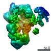


 Keywords
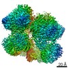


 enterobacteria phage t7 (virus)
