[English] 日本語
Yorodumi
- PDB-1t8e: T7 DNA Polymerase Ternary Complex with dCTP at the Insertion Site. -

+
Open data


ID or keywords:

Loading...

-
Basic information

Entry
Database: PDB / ID: 1t8e
TitleT7 DNA Polymerase Ternary Complex with dCTP at the Insertion Site.
Components
  • 25-MER
  • 5'-D(P*CP*GP*AP*AP*AP*AP*CP*GP*AP*CP*GP*GP*CP*CP*AP*GP*TP*GP*CP*CP*AP*(2DT))-3'
  • DNA polymerase
  • thioredoxin 1
Keywords
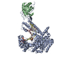
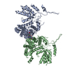
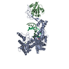
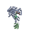
transferase/electron transport/DNA / protein / DNA / transferase / transferase-electron transport-DNA COMPLEX
Function / homology
Function and homology information


DNA synthesis involved in DNA replication / DNA exonuclease activity / viral DNA genome replication / Hydrolases; Acting on ester bonds; Exodeoxyribonucleases producing 5'-phosphomonoesters / DNA polymerase processivity factor activity / protein-disulfide reductase activity / 3'-5' exonuclease activity / cell redox homeostasis / DNA-templated DNA replication / DNA-directed DNA polymerase ...DNA synthesis involved in DNA replication / DNA exonuclease activity / viral DNA genome replication / Hydrolases; Acting on ester bonds; Exodeoxyribonucleases producing 5'-phosphomonoesters / DNA polymerase processivity factor activity / protein-disulfide reductase activity / 3'-5' exonuclease activity / cell redox homeostasis / DNA-templated DNA replication / DNA-directed DNA polymerase / DNA-directed DNA polymerase activity / nucleotide binding / DNA binding / metal ion binding / cytosol / cytoplasm
Similarity search - Function
DNA-directed DNA polymerase T7 / Taq DNA Polymerase; Chain T, domain 4 / Taq DNA Polymerase; Chain T, domain 4 / Alpha-Beta Plaits - #370 / Thioredoxin / DNA polymerase A / DNA polymerase family A / DNA-directed DNA polymerase, family A, conserved site / DNA polymerase family A signature. / DNA-directed DNA polymerase, family A, palm domain ...DNA-directed DNA polymerase T7 / Taq DNA Polymerase; Chain T, domain 4 / Taq DNA Polymerase; Chain T, domain 4 / Alpha-Beta Plaits - #370 / Thioredoxin / DNA polymerase A / DNA polymerase family A / DNA-directed DNA polymerase, family A, conserved site / DNA polymerase family A signature. / DNA-directed DNA polymerase, family A, palm domain / DNA polymerase A domain / Thioredoxin / Thioredoxin, conserved site / Thioredoxin family active site. / Thioredoxin domain profile. / Thioredoxin domain / 5' to 3' exonuclease, C-terminal subdomain / Ribonuclease H-like superfamily/Ribonuclease H / Glutaredoxin / Glutaredoxin / DNA polymerase; domain 1 / Nucleotidyltransferase; domain 5 / Thioredoxin-like superfamily / Ribonuclease H superfamily / Ribonuclease H-like superfamily / Alpha-Beta Plaits / DNA/RNA polymerase superfamily / Up-down Bundle / 2-Layer Sandwich / Orthogonal Bundle / 3-Layer(aba) Sandwich / Mainly Alpha / Alpha Beta
Similarity search - Domain/homology
2',3'-DIDEOXYCYTIDINE 5'-TRIPHOSPHATE / DNA / DNA (> 10) / DNA-directed DNA polymerase / Thioredoxin 1
Similarity search - Component
Biological speciesEnterobacteria phage T7 (virus)
Escherichia coli (E. coli)
MethodX-RAY DIFFRACTION / MOLECULAR REPLACEMENT / Resolution: 2.54 Å
AuthorsBrieba, L.G. / Eichman, B.F. / Kokoska, R.J. / Doublie, S. / Kunkel, T.A. / Ellenberger, T.
CitationJournal: Embo J. / Year: 2004
Title: Structural basis for the dual coding potential of 8-oxoguanosine by a high-fidelity DNA polymerase.
Authors: Brieba, L.G. / Eichman, B.F. / Kokoska, R.J. / Doublie, S. / Kunkel, T.A. / Ellenberger, T.
History
DepositionMay 12, 2004Deposition site: RCSB / Processing site: RCSB
Revision 1.0Oct 12, 2004Provider: repository / Type: Initial release
Revision 1.1Apr 30, 2008Group: Version format compliance
Revision 1.2Jul 13, 2011Group: Version format compliance
Revision 1.3Feb 14, 2024Group: Data collection / Database references / Derived calculations
Category: chem_comp_atom / chem_comp_bond ...chem_comp_atom / chem_comp_bond / database_2 / pdbx_struct_conn_angle / struct_conn / struct_conn_type / struct_ref_seq_dif / struct_site
Item: _database_2.pdbx_DOI / _database_2.pdbx_database_accession ..._database_2.pdbx_DOI / _database_2.pdbx_database_accession / _pdbx_struct_conn_angle.ptnr1_auth_comp_id / _pdbx_struct_conn_angle.ptnr1_auth_seq_id / _pdbx_struct_conn_angle.ptnr1_label_asym_id / _pdbx_struct_conn_angle.ptnr1_label_atom_id / _pdbx_struct_conn_angle.ptnr1_label_comp_id / _pdbx_struct_conn_angle.ptnr1_label_seq_id / _pdbx_struct_conn_angle.ptnr2_auth_seq_id / _pdbx_struct_conn_angle.ptnr2_label_asym_id / _pdbx_struct_conn_angle.ptnr3_auth_comp_id / _pdbx_struct_conn_angle.ptnr3_auth_seq_id / _pdbx_struct_conn_angle.ptnr3_label_asym_id / _pdbx_struct_conn_angle.ptnr3_label_atom_id / _pdbx_struct_conn_angle.ptnr3_label_comp_id / _pdbx_struct_conn_angle.ptnr3_label_seq_id / _pdbx_struct_conn_angle.value / _struct_conn.conn_type_id / _struct_conn.id / _struct_conn.pdbx_dist_value / _struct_conn.pdbx_leaving_atom_flag / _struct_conn.ptnr1_auth_asym_id / _struct_conn.ptnr1_auth_comp_id / _struct_conn.ptnr1_auth_seq_id / _struct_conn.ptnr1_label_asym_id / _struct_conn.ptnr1_label_atom_id / _struct_conn.ptnr1_label_comp_id / _struct_conn.ptnr1_label_seq_id / _struct_conn.ptnr2_auth_asym_id / _struct_conn.ptnr2_auth_comp_id / _struct_conn.ptnr2_auth_seq_id / _struct_conn.ptnr2_label_asym_id / _struct_conn.ptnr2_label_atom_id / _struct_conn.ptnr2_label_comp_id / _struct_conn.ptnr2_label_seq_id / _struct_conn_type.id / _struct_ref_seq_dif.details / _struct_site.pdbx_auth_asym_id / _struct_site.pdbx_auth_comp_id / _struct_site.pdbx_auth_seq_id

-
Structure visualization

Structure viewerMolecule:
MolmilJmol/JSmol

Downloads & links

-
Assembly

Deposited unit
C: 5'-D(P*CP*GP*AP*AP*AP*AP*CP*GP*AP*CP*GP*GP*CP*CP*AP*GP*TP*GP*CP*CP*AP*(2DT))-3'
D: 25-MER
A: DNA polymerase
B: thioredoxin 1
hetero molecules


Theoretical massNumber of molelcules
Total (without water)106,55211
Polymers105,5434
Non-polymers1,0107
Water6,864381
1


  • Idetical with deposited unit
  • defined by author
TypeNameSymmetry operationNumber
identity operation1_555x,y,z1
Unit cell
Length a, b, c (Å)106.285, 218.121, 52.133
Angle α, β, γ (deg.)90.00, 90.00, 90.00
Int Tables number18
Space group name H-MP21212

-
Components

-
DNA chain , 2 types, 2 molecules CD

#1: DNA chain 5'-D(P*CP*GP*AP*AP*AP*AP*CP*GP*AP*CP*GP*GP*CP*CP*AP*GP*TP*GP*CP*CP*AP*(2DT))-3'


Mass: 6739.384 Da / Num. of mol.: 1 / Source method: obtained synthetically
#2: DNA chain 25-MER


Mass: 8026.148 Da / Num. of mol.: 1 / Source method: obtained synthetically

-
Protein , 2 types, 2 molecules AB

#3: Protein DNA polymerase / / T7 DNA polymerase


Mass: 79089.789 Da / Num. of mol.: 1
Source method: isolated from a genetically manipulated source
Source: (gene. exp.) Enterobacteria phage T7 (virus) / Genus: T7-like viruses / Gene: 5, T7DNApolymerase / Plasmid: pGP5 / Species (production host): Escherichia coli / Production host: Escherichia coli BL21(DE3) (bacteria) / Strain (production host): BL21DE3 / References: UniProt: P00581, DNA-directed DNA polymerase
#4: Protein thioredoxin 1 /


Mass: 11687.388 Da / Num. of mol.: 1
Source method: isolated from a genetically manipulated source
Source: (gene. exp.) Escherichia coli (E. coli) / Gene: trxa, tsnc, fipa, b3781, c4701, z5291,Thioredoxin / Plasmid: pTrx-3 / Species (production host): Escherichia coli / Production host: Escherichia coli BL21(DE3) (bacteria) / Strain (production host): BL21DE3 / References: UniProt: P0AA25

-
Non-polymers , 6 types, 388 molecules

#5: Chemical ChemComp-MG / MAGNESIUM ION


Mass: 24.305 Da / Num. of mol.: 3 / Source method: obtained synthetically / Formula: Mg
#6: Chemical ChemComp-SO4 / SULFATE ION / Sulfate


Mass: 96.063 Da / Num. of mol.: 1 / Source method: obtained synthetically / Formula: SO4
#7: Chemical ChemComp-DCT / 2',3'-DIDEOXYCYTIDINE 5'-TRIPHOSPHATE


Type: DNA linking / Mass: 451.158 Da / Num. of mol.: 1 / Source method: obtained synthetically / Formula: C9H16N3O12P3
#8: Chemical ChemComp-MES / 2-(N-MORPHOLINO)-ETHANESULFONIC ACID / MES (buffer)


Mass: 195.237 Da / Num. of mol.: 1 / Source method: obtained synthetically / Formula: C6H13NO4S / Comment: pH buffer*YM
#9: Chemical ChemComp-PG4 / TETRAETHYLENE GLYCOL / Polyethylene glycol


Mass: 194.226 Da / Num. of mol.: 1 / Source method: obtained synthetically / Formula: C8H18O5 / Comment: precipitant*YM
#10: Water ChemComp-HOH / water / Water


Mass: 18.015 Da / Num. of mol.: 381 / Source method: isolated from a natural source / Formula: H2O

-
Experimental details

-
Experiment

ExperimentMethod: X-RAY DIFFRACTION / Number of used crystals: 1

-
Sample preparation

CrystalDensity Matthews: 2.85 Å3/Da / Density % sol: 56.77 %

-
Data collection

DetectorDate: Dec 10, 1998
RadiationProtocol: SINGLE WAVELENGTH / Monochromatic (M) / Laue (L): M / Scattering type: x-ray
Radiation wavelengthRelative weight: 1
ReflectionResolution: 2.54→40 Å / Num. obs: 39002 / Biso Wilson estimate: 37.1 Å2

-
Processing

Software
NameVersionClassification
CNS1.1refinement
HKL-2000data reduction
SCALEPACKdata scaling
AMoREphasing
RefinementMethod to determine structure: MOLECULAR REPLACEMENT / Resolution: 2.54→28.71 Å / Rfactor Rfree error: 0.006 / Data cutoff high absF: 1566872.8 / Data cutoff high rms absF: 1566872.8 / Data cutoff low absF: 0 / Isotropic thermal model: RESTRAINED / Cross valid method: THROUGHOUT / σ(F): 0 / Stereochemistry target values: Engh & Huber
RfactorNum. reflection% reflectionSelection details
Rfree0.255 1922 4.9 %RANDOM
Rwork0.22 ---
all0.255 ---
obs0.22 37002 95 %-
Solvent computationSolvent model: FLAT MODEL / Bsol: 30.7431 Å2 / ksol: 0.309425 e/Å3
Displacement parametersBiso mean: 39.2 Å2
Baniso -1Baniso -2Baniso -3
1-1.33 Å20 Å20 Å2
2--0.14 Å20 Å2
3----1.47 Å2
Refine analyze
FreeObs
Luzzati coordinate error0.35 Å0.31 Å
Luzzati d res low-5 Å
Luzzati sigma a0.4 Å0.32 Å
Refinement stepCycle: LAST / Resolution: 2.54→28.71 Å
ProteinNucleic acidLigandSolventTotal
Num. atoms6335 965 60 381 7741
Refine LS restraints
Refine-IDTypeDev idealDev ideal target
X-RAY DIFFRACTIONc_bond_d0.006
X-RAY DIFFRACTIONc_bond_d_na
X-RAY DIFFRACTIONc_bond_d_prot
X-RAY DIFFRACTIONc_angle_d
X-RAY DIFFRACTIONc_angle_d_na
X-RAY DIFFRACTIONc_angle_d_prot
X-RAY DIFFRACTIONc_angle_deg1.2
X-RAY DIFFRACTIONc_angle_deg_na
X-RAY DIFFRACTIONc_angle_deg_prot
X-RAY DIFFRACTIONc_dihedral_angle_d21.5
X-RAY DIFFRACTIONc_dihedral_angle_d_na
X-RAY DIFFRACTIONc_dihedral_angle_d_prot
X-RAY DIFFRACTIONc_improper_angle_d0.98
X-RAY DIFFRACTIONc_improper_angle_d_na
X-RAY DIFFRACTIONc_improper_angle_d_prot
X-RAY DIFFRACTIONc_mcbond_it1.181.5
X-RAY DIFFRACTIONc_mcangle_it1.972
X-RAY DIFFRACTIONc_scbond_it1.722
X-RAY DIFFRACTIONc_scangle_it2.532.5
LS refinement shellResolution: 2.54→2.7 Å / Rfactor Rfree error: 0.02 / Total num. of bins used: 6
RfactorNum. reflection% reflection
Rfree0.34 303 4.9 %
Rwork0.3 5856 -
obs--91.7 %
Xplor file
Refine-IDSerial noParam fileTopol file
X-RAY DIFFRACTION1PROTEIN_REP.PARAMPROTEIN.TOP
X-RAY DIFFRACTION2DNA-RNA-TT.PARAMDNA-RNA-TT.TOP
X-RAY DIFFRACTION3ION.PARAMION.TOP
X-RAY DIFFRACTION4WATER.PARAMWATER.TOP
X-RAY DIFFRACTION5LIGAND.PARLIGAND.TOP

+
About Yorodumi

-
News

-
Feb 9, 2022. New format data for meta-information of EMDB entries

New format data for meta-information of EMDB entries

  • Version 3 of the EMDB header file is now the official format.
  • The previous official version 1.9 will be removed from the archive.

Related info.:EMDB header

External links:wwPDB to switch to version 3 of the EMDB data model

-
Aug 12, 2020. Covid-19 info

Covid-19 info

URL: https://pdbj.org/emnavi/covid19.php

New page: Covid-19 featured information page in EM Navigator.

Related info.:Covid-19 info / Mar 5, 2020. Novel coronavirus structure data

+
Mar 5, 2020. Novel coronavirus structure data

Novel coronavirus structure data

Related info.:Yorodumi Speices / Aug 12, 2020. Covid-19 info

External links:COVID-19 featured content - PDBj / Molecule of the Month (242):Coronavirus Proteases

+
Jan 31, 2019. EMDB accession codes are about to change! (news from PDBe EMDB page)

EMDB accession codes are about to change! (news from PDBe EMDB page)

  • The allocation of 4 digits for EMDB accession codes will soon come to an end. Whilst these codes will remain in use, new EMDB accession codes will include an additional digit and will expand incrementally as the available range of codes is exhausted. The current 4-digit format prefixed with “EMD-” (i.e. EMD-XXXX) will advance to a 5-digit format (i.e. EMD-XXXXX), and so on. It is currently estimated that the 4-digit codes will be depleted around Spring 2019, at which point the 5-digit format will come into force.
  • The EM Navigator/Yorodumi systems omit the EMD- prefix.

Related info.:Q: What is EMD? / ID/Accession-code notation in Yorodumi/EM Navigator

External links:EMDB Accession Codes are Changing Soon! / Contact to PDBj

+
Jul 12, 2017. Major update of PDB

Major update of PDB

  • wwPDB released updated PDB data conforming to the new PDBx/mmCIF dictionary.
  • This is a major update changing the version number from 4 to 5, and with Remediation, in which all the entries are updated.
  • In this update, many items about electron microscopy experimental information are reorganized (e.g. em_software).
  • Now, EM Navigator and Yorodumi are based on the updated data.

External links:wwPDB Remediation / Enriched Model Files Conforming to OneDep Data Standards Now Available in the PDB FTP Archive

-
Yorodumi

Thousand views of thousand structures

  • Yorodumi is a browser for structure data from EMDB, PDB, SASBDB, etc.
  • This page is also the successor to EM Navigator detail page, and also detail information page/front-end page for Omokage search.
  • The word "yorodu" (or yorozu) is an old Japanese word meaning "ten thousand". "mi" (miru) is to see.

Related info.:EMDB / PDB / SASBDB / Comparison of 3 databanks / Yorodumi Search / Aug 31, 2016. New EM Navigator & Yorodumi / Yorodumi Papers / Jmol/JSmol / Function and homology information / Changes in new EM Navigator and Yorodumi

Read more