DNA synthesis involved in DNA replication / DNA exonuclease activity / viral DNA genome replication / Hydrolases; Acting on ester bonds; Exodeoxyribonucleases producing 5'-phosphomonoesters / DNA polymerase processivity factor activity / protein-disulfide reductase activity / 3'-5' exonuclease activity / cell redox homeostasis / DNA-templated DNA replication / double-strand break repair ...DNA synthesis involved in DNA replication / DNA exonuclease activity / viral DNA genome replication / Hydrolases; Acting on ester bonds; Exodeoxyribonucleases producing 5'-phosphomonoesters / DNA polymerase processivity factor activity / protein-disulfide reductase activity / 3'-5' exonuclease activity / cell redox homeostasis / DNA-templated DNA replication / double-strand break repair / DNA-directed DNA polymerase / DNA-directed DNA polymerase activity / nucleotide binding / DNA binding / metal ion binding / cytoplasm / cytosol Similarity search - Function
DNA-directed DNA polymerase T7 / Taq DNA Polymerase; Chain T, domain 4 / Taq DNA Polymerase; Chain T, domain 4 / Alpha-Beta Plaits - #370 / Thioredoxin / DNA polymerase A / DNA polymerase family A / DNA-directed DNA polymerase, family A, conserved site / DNA polymerase family A signature. / DNA-directed DNA polymerase, family A, palm domain ...DNA-directed DNA polymerase T7 / Taq DNA Polymerase; Chain T, domain 4 / Taq DNA Polymerase; Chain T, domain 4 / Alpha-Beta Plaits - #370 / Thioredoxin / DNA polymerase A / DNA polymerase family A / DNA-directed DNA polymerase, family A, conserved site / DNA polymerase family A signature. / DNA-directed DNA polymerase, family A, palm domain / DNA polymerase A domain / Thioredoxin / Thioredoxin family active site. / Thioredoxin, conserved site / Thioredoxin domain profile. / Thioredoxin domain / 5' to 3' exonuclease, C-terminal subdomain / Ribonuclease H-like superfamily/Ribonuclease H / Glutaredoxin / Glutaredoxin / DNA polymerase; domain 1 / Nucleotidyltransferase; domain 5 / Thioredoxin-like superfamily / Ribonuclease H superfamily / Ribonuclease H-like superfamily / Alpha-Beta Plaits / DNA/RNA polymerase superfamily / Up-down Bundle / 2-Layer Sandwich / Orthogonal Bundle / 3-Layer(aba) Sandwich / Mainly Alpha / Alpha Beta Similarity search - Domain/homology
Mass: 6739.384 Da / Num. of mol.: 2 / Source method: obtained synthetically
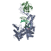
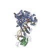
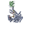
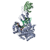
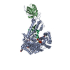
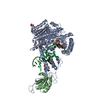
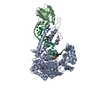
#2: DNA chain
DNATemplate
Mass: 8026.148 Da / Num. of mol.: 2 / Source method: obtained synthetically
#3: Protein
T7DNApolymerase / E.C.2.7.7.7
Mass: 79703.578 Da / Num. of mol.: 2 / Mutation: Residues 5 and 7 mutated to Ala Source method: isolated from a genetically manipulated source Source: (gene. exp.) Enterobacteria phage T7 (virus) / Genus: T7-like viruses Description: Protein induced at an OD_600 of 0.5 by addition of 0.5 mM IPTG for 4 hours at 37C Gene: 5 / Plasmid: pGP5 / Production host: Escherichia coli (E. coli) / Strain (production host): BL21 (DE3) pLys S / References: UniProt: P00581, DNA-directed DNA polymerase
#4: Protein
thioredoxin1 / TRX1 / TRX
Mass: 11687.388 Da / Num. of mol.: 2 / Mutation: Residues 5 and 7 mutated to Ala Source method: isolated from a genetically manipulated source Source: (gene. exp.) Escherichia coli (E. coli) Description: Protein induced at an OD_600 of 0.5 by addition of 0.5 mM IPTG for 4 hours at 37C Gene: trxA, fipA, tsnC / Plasmid: p-Trx / Production host: Escherichia coli (E. coli) / Strain (production host): BL21 (DE3) pLys S / References: UniProt: P0AA25
Mass: 18.015 Da / Num. of mol.: 414 / Source method: isolated from a natural source / Formula: H2O
-
Experimental details
-
Experiment
Experiment
Method: X-RAY DIFFRACTION / Number of used crystals: 1
-
Sample preparation
Crystal
Density Matthews: 3.18 Å3/Da / Density % sol: 61 %
Crystal grow
Temperature: 273 K / Method: vapor diffusion, hanging drop / pH: 7.5 Details: A complex of 1x10^-4 M T7 DNA polymerase 5A7A:thioredoxin was assembled with an equimolar amount of double stranded DNA substrate. Crystallization was achieved using a buffer containing 50mM ...Details: A complex of 1x10^-4 M T7 DNA polymerase 5A7A:thioredoxin was assembled with an equimolar amount of double stranded DNA substrate. Crystallization was achieved using a buffer containing 50mM HEPES pH 7.5, 10mM MgCl_2, 2mM DTT, and 0.5 mM terminal ddTTP Seed crystals were grown by hanging drop vapor diffusion by mixing 1ul each of protein-DNA solution and a reservoir solutions containing between 16 to 20% PEG 8000, 100mM ACES pH 7.5, 120 ammonium sulfate, 30mM MgCl2, and 5mM DTT. These crystals were used to streak-seed a grid of protein/reservoir solutions with concentrations of PEG 8000 between 13 to 15%. Pyramidal crystals appeared overnight and reached a maximum size of ~150 X 150 X 100 um3 after 3 to 4 days. Crystals were harvested overnight in mother-liquor containing 10 % PEG 400, temperature 273K, VAPOR DIFFUSION, HANGING DROP
In the structure databanks used in Yorodumi, some data are registered as the other names, "COVID-19 virus" and "2019-nCoV". Here are the details of the virus and the list of structure data.
Jan 31, 2019. EMDB accession codes are about to change! (news from PDBe EMDB page)
EMDB accession codes are about to change! (news from PDBe EMDB page)
The allocation of 4 digits for EMDB accession codes will soon come to an end. Whilst these codes will remain in use, new EMDB accession codes will include an additional digit and will expand incrementally as the available range of codes is exhausted. The current 4-digit format prefixed with “EMD-” (i.e. EMD-XXXX) will advance to a 5-digit format (i.e. EMD-XXXXX), and so on. It is currently estimated that the 4-digit codes will be depleted around Spring 2019, at which point the 5-digit format will come into force.
The EM Navigator/Yorodumi systems omit the EMD- prefix.
Related info.:Q: What is EMD? / ID/Accession-code notation in Yorodumi/EM Navigator
Yorodumi is a browser for structure data from EMDB, PDB, SASBDB, etc.
This page is also the successor to EM Navigator detail page, and also detail information page/front-end page for Omokage search.
The word "yorodu" (or yorozu) is an old Japanese word meaning "ten thousand". "mi" (miru) is to see.
Related info.:EMDB / PDB / SASBDB / Comparison of 3 databanks / Yorodumi Search / Aug 31, 2016. New EM Navigator & Yorodumi / Yorodumi Papers / Jmol/JSmol / Function and homology information / Changes in new EM Navigator and Yorodumi
 Movie
Movie Controller

 Yorodumi
 Open data
 Basic information
 Components
 Keywords
 Function and homology information
 Enterobacteria phage T7 (virus)
 X-RAY DIFFRACTION /
  Authors
 Citation
 Structure visualization
 Downloads & links
 Other downloads


 PDBj
PDBj


 Assembly

 Mass: 18.015 Da / Num. of mol.: 414 / Source method: isolated from a natural source / Formula: H2O
 Sample preparation
 / Beamline: X26C / Wavelength: 1.1 / Wavelength: 1.1 Å
 Processing