 Movie
Movie Controller

[English] 日本語
 Yorodumi
- PDB-6g0p: Crystal Structure of the first bromodomain of human BRD4 in compl... -
+ Open data

- Basic information
| Entry | Database: PDB / ID: 6g0p | ||||||
|---|---|---|---|---|---|---|---|
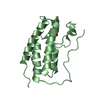
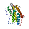
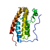
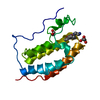
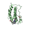
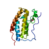
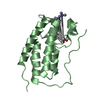
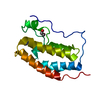
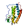
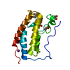
| Title | Crystal Structure of the first bromodomain of human BRD4 in complex with an acetylated E2F1 peptide (K117ac/K120ac) | ||||||
 Components Components |
| ||||||
 Keywords Keywords | TRANSCRIPTION / Bromodomain / complex | ||||||
| Function / homology |  Function and homology information Function and homology informationlens fiber cell apoptotic process / Rb-E2F complex / negative regulation of fat cell proliferation / negative regulation of DNA binding / Inhibition of replication initiation of damaged DNA by RB1/E2F1 / quinolinate biosynthetic process / Transcription of E2F targets under negative control by p107 (RBL1) and p130 (RBL2) in complex with HDAC1 / cellular response to fatty acid / Transcription of E2F targets under negative control by DREAM complex / Activation of NOXA and translocation to mitochondria ...lens fiber cell apoptotic process / Rb-E2F complex / negative regulation of fat cell proliferation / negative regulation of DNA binding / Inhibition of replication initiation of damaged DNA by RB1/E2F1 / quinolinate biosynthetic process / Transcription of E2F targets under negative control by p107 (RBL1) and p130 (RBL2) in complex with HDAC1 / cellular response to fatty acid / Transcription of E2F targets under negative control by DREAM complex / Activation of NOXA and translocation to mitochondria / anoikis / Activation of PUMA and translocation to mitochondria / mRNA stabilization / forebrain development / DNA-binding transcription activator activity / negative regulation of fat cell differentiation / G2 Phase / G1/S-Specific Transcription / nuclear chromosome / histone H4K8ac reader activity / Defective binding of RB1 mutants to E2F1,(E2F2, E2F3) / RNA polymerase II C-terminal domain binding / histone H3K27ac reader activity / Transcriptional Regulation by E2F6 / P-TEFb complex binding / histone H3K9ac reader activity / negative regulation of DNA damage checkpoint / intrinsic apoptotic signaling pathway by p53 class mediator / histone H4 reader activity / histone H4K5ac reader activity / histone H4K12ac reader activity / host-mediated suppression of viral transcription / histone H4K16ac reader activity / positive regulation of glial cell proliferation / positive regulation of G2/M transition of mitotic cell cycle / TP53 Regulates Transcription of Genes Involved in G1 Cell Cycle Arrest / positive regulation of T-helper 17 cell lineage commitment / Cyclin E associated events during G1/S transition / Cyclin A:Cdk2-associated events at S phase entry / cis-regulatory region sequence-specific DNA binding / regulation of G1/S transition of mitotic cell cycle / RNA polymerase II CTD heptapeptide repeat kinase activity / DNA damage checkpoint signaling / condensed nuclear chromosome / transcription coregulator activity / positive regulation of transcription elongation by RNA polymerase II / cellular response to nerve growth factor stimulus / cellular response to xenobiotic stimulus / Oncogene Induced Senescence / intrinsic apoptotic signaling pathway in response to DNA damage / Pre-NOTCH Transcription and Translation / RNA polymerase II transcription regulator complex / positive regulation of fibroblast proliferation / Transcriptional regulation of granulopoiesis / sequence-specific double-stranded DNA binding / p53 binding / Cyclin D associated events in G1 / chromosome / regulation of inflammatory response / response to lipopolysaccharide / histone binding / Oxidative Stress Induced Senescence / cellular response to hypoxia / DNA-binding transcription factor binding / spermatogenesis / sequence-specific DNA binding / Potential therapeutics for SARS / molecular adaptor activity / DNA-binding transcription factor activity, RNA polymerase II-specific / positive regulation of canonical NF-kappaB signal transduction / transcription coactivator activity / protein dimerization activity / transcription cis-regulatory region binding / RNA polymerase II cis-regulatory region sequence-specific DNA binding / positive regulation of apoptotic process / chromatin remodeling / DNA-binding transcription factor activity / negative regulation of DNA-templated transcription / protein serine/threonine kinase activity / chromatin binding / centrosome / positive regulation of gene expression / regulation of transcription by RNA polymerase II / regulation of DNA-templated transcription / DNA damage response / protein kinase binding / positive regulation of DNA-templated transcription / chromatin / enzyme binding / negative regulation of transcription by RNA polymerase II / DNA-templated transcription / positive regulation of transcription by RNA polymerase II / protein-containing complex / DNA binding / nucleoplasm / nucleus / cytoplasm Similarity search - Function | ||||||
| Biological species |  Homo sapiens (human) Homo sapiens (human) | ||||||
| Method |  X-RAY DIFFRACTION / SYNCHROTRON / MOLECULAR REPLACEMENT / molecular replacement / Resolution: 1.3 Å X-RAY DIFFRACTION / SYNCHROTRON / MOLECULAR REPLACEMENT / molecular replacement / Resolution: 1.3 Å | ||||||
 Authors Authors | Filippakopoulos, P. / Picaud, S. / Krojer, T. / Sorrell, F. / Pike, A.C.W. / von Delft, F. / Arrowsmith, C.H. / Edwards, A.M. / Bountra, C. | ||||||
| Funding support |  United Kingdom, 1items United Kingdom, 1items
| ||||||
 Citation Citation | Journal: Mol Cell / Year: 2019 Title: Interactome Rewiring Following Pharmacological Targeting of BET Bromodomains. Authors: Jean-Philippe Lambert / Sarah Picaud / Takao Fujisawa / Huayun Hou / Pavel Savitsky / Liis Uusküla-Reimand / Gagan D Gupta / Hala Abdouni / Zhen-Yuan Lin / Monika Tucholska / James D R ...Authors: Jean-Philippe Lambert / Sarah Picaud / Takao Fujisawa / Huayun Hou / Pavel Savitsky / Liis Uusküla-Reimand / Gagan D Gupta / Hala Abdouni / Zhen-Yuan Lin / Monika Tucholska / James D R Knight / Beatriz Gonzalez-Badillo / Nicole St-Denis / Joseph A Newman / Manuel Stucki / Laurence Pelletier / Nuno Bandeira / Michael D Wilson / Panagis Filippakopoulos / Anne-Claude Gingras /     Abstract: Targeting bromodomains (BRDs) of the bromo-and-extra-terminal (BET) family offers opportunities for therapeutic intervention in cancer and other diseases. Here, we profile the interactomes of BRD2, ...Targeting bromodomains (BRDs) of the bromo-and-extra-terminal (BET) family offers opportunities for therapeutic intervention in cancer and other diseases. Here, we profile the interactomes of BRD2, BRD3, BRD4, and BRDT following treatment with the pan-BET BRD inhibitor JQ1, revealing broad rewiring of the interaction landscape, with three distinct classes of behavior for the 603 unique interactors identified. A group of proteins associate in a JQ1-sensitive manner with BET BRDs through canonical and new binding modes, while two classes of extra-terminal (ET)-domain binding motifs mediate acetylation-independent interactions. Last, we identify an unexpected increase in several interactions following JQ1 treatment that define negative functions for BRD3 in the regulation of rRNA synthesis and potentially RNAPII-dependent gene expression that result in decreased cell proliferation. Together, our data highlight the contributions of BET protein modules to their interactomes allowing for a better understanding of pharmacological rewiring in response to JQ1. | ||||||
| History |
|
- Structure visualization
| Structure viewer | Molecule: MolmilJmol/JSmol |
|---|
- Downloads & links
-Download
| PDBx/mmCIF format | 6g0p.cif.gz | 79 KB | Display | PDBx/mmCIF format |
|---|---|---|---|---|
| PDB format | pdb6g0p.ent.gz | 56.9 KB | Display | PDB format |
| PDBx/mmJSON format | 6g0p.json.gz | Tree view | PDBx/mmJSON format | |
| Others |  Other downloads Other downloads |
-Validation report
| Arichive directory | https://data.pdbj.org/pub/pdb/validation_reports/g0/6g0pftp://data.pdbj.org/pub/pdb/validation_reports/g0/6g0p | HTTPS FTP |
|---|
-Related structure data
| Related structure data | 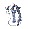 5nncC 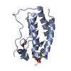 5nndC  5nneC  5nnfC  5nngC  6g0oC  6g0qC  6g0rC 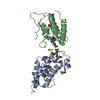 6g0sC 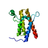 2grcS 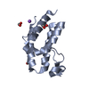 2oo1S 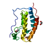 2ossS 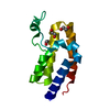 2ouoS 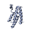 3d7cS 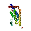 3daiS  3dwyS S: Starting model for refinement C: citing same article ( |
|---|---|
| Similar structure data |


-Links
 PDBj
PDBj


- Assembly
| Deposited unit | 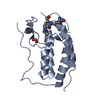
| ||||||||
|---|---|---|---|---|---|---|---|---|---|
| 1 |
| ||||||||
| Unit cell |
|
-Components
| #1: Protein | Mass: 15099.380 Da / Num. of mol.: 1 Source method: isolated from a genetically manipulated source Source: (gene. exp.) Homo sapiens (human) / Gene: BRD4, HUNK1 / Plasmid: pNIC28-Bsa4 / Production host:  |
|---|---|
| #2: Protein/peptide | Mass: 1843.026 Da / Num. of mol.: 1 / Source method: obtained synthetically / Details: E2F1 peptide acetylated at K117 and K120 / Source: (synth.) Homo sapiens (human) / References: UniProt: Q01094 |
| #3: Chemical | ChemComp-EDO /   Mass: 62.068 Da / Num. of mol.: 1 / Source method: obtained synthetically / Formula: C2H6O2 Mass: 62.068 Da / Num. of mol.: 1 / Source method: obtained synthetically / Formula: C2H6O2 |
| #4: Water | ChemComp-HOH /  Mass: 18.015 Da / Num. of mol.: 174 / Source method: isolated from a natural source / Formula: H2O Mass: 18.015 Da / Num. of mol.: 174 / Source method: isolated from a natural source / Formula: H2O |
| Has protein modification | Y |
-Experimental details
-Experiment
| Experiment | Method: X-RAY DIFFRACTION / Number of used crystals: 1 |
|---|
- Sample preparation
| Crystal | Density Matthews: 2.16 Å3/Da / Density % sol: 42.92 % |
|---|---|
| Crystal grow | Temperature: 277 K / Method: vapor diffusion, sitting drop / pH: 7 / Details: 20.0 % PEG3350 10.0 % EtGly 0.2 M NaCHO |
-Data collection
| Diffraction | Mean temperature: 100 K | |||||||||||||||||||||||||||||||||||||||||||||||||||||||||||||||||||||||||||||||||||||||||||||||||||
|---|---|---|---|---|---|---|---|---|---|---|---|---|---|---|---|---|---|---|---|---|---|---|---|---|---|---|---|---|---|---|---|---|---|---|---|---|---|---|---|---|---|---|---|---|---|---|---|---|---|---|---|---|---|---|---|---|---|---|---|---|---|---|---|---|---|---|---|---|---|---|---|---|---|---|---|---|---|---|---|---|---|---|---|---|---|---|---|---|---|---|---|---|---|---|---|---|---|---|---|---|
| Diffraction source | Source: SYNCHROTRON / Site: Diamond / Beamline: I03 / Wavelength: 0.9763 Å | |||||||||||||||||||||||||||||||||||||||||||||||||||||||||||||||||||||||||||||||||||||||||||||||||||
| Detector | Type: DECTRIS PILATUS 6M-F / Detector: PIXEL / Date: Dec 13, 2015 | |||||||||||||||||||||||||||||||||||||||||||||||||||||||||||||||||||||||||||||||||||||||||||||||||||
| Radiation | Protocol: SINGLE WAVELENGTH / Monochromatic (M) / Laue (L): M / Scattering type: x-ray | |||||||||||||||||||||||||||||||||||||||||||||||||||||||||||||||||||||||||||||||||||||||||||||||||||
| Radiation wavelength | Wavelength: 0.9763 Å / Relative weight: 1 | |||||||||||||||||||||||||||||||||||||||||||||||||||||||||||||||||||||||||||||||||||||||||||||||||||
| Reflection | Resolution: 1.3→58.126 Å / Num. all: 34488 / Num. obs: 34488 / % possible obs: 99.9 % / Redundancy: 6.7 % / Rpim(I) all: 0.014 / Rrim(I) all: 0.036 / Rsym value: 0.034 / Net I/av σ(I): 9.6 / Net I/σ(I): 29.6 / Num. measured all: 232698 | |||||||||||||||||||||||||||||||||||||||||||||||||||||||||||||||||||||||||||||||||||||||||||||||||||
| Reflection shell | Diffraction-ID: 1
|
-Phasing
| Phasing | Method: molecular replacement | |||||||||
|---|---|---|---|---|---|---|---|---|---|---|
| Phasing MR | Model details: Phaser MODE: MR_AUTO
|
- Processing
| Software |
| |||||||||||||||||||||||||||||||||||||||||||||||||||||||||||||||||||||||||||
|---|---|---|---|---|---|---|---|---|---|---|---|---|---|---|---|---|---|---|---|---|---|---|---|---|---|---|---|---|---|---|---|---|---|---|---|---|---|---|---|---|---|---|---|---|---|---|---|---|---|---|---|---|---|---|---|---|---|---|---|---|---|---|---|---|---|---|---|---|---|---|---|---|---|---|---|---|
| Refinement | Method to determine structure: MOLECULAR REPLACEMENT Starting model: Ensemble of 2OSS, 2OUO, 2GRC, 2OO1, 3DAI, 3D7C, 3DWY Resolution: 1.3→39.31 Å / Cor.coef. Fo:Fc: 0.979 / Cor.coef. Fo:Fc free: 0.976 / SU B: 1.061 / SU ML: 0.021 / SU R Cruickshank DPI: 0.0413 / Cross valid method: THROUGHOUT / σ(F): 0 / ESU R: 0.041 / ESU R Free: 0.039 Details: HYDROGENS HAVE BEEN ADDED IN THE RIDING POSITIONS U VALUES : REFINED INDIVIDUALLY
| |||||||||||||||||||||||||||||||||||||||||||||||||||||||||||||||||||||||||||
| Solvent computation | Ion probe radii: 0.8 Å / Shrinkage radii: 0.8 Å / VDW probe radii: 1.2 Å | |||||||||||||||||||||||||||||||||||||||||||||||||||||||||||||||||||||||||||
| Displacement parameters | Biso max: 62.72 Å2 / Biso mean: 16.906 Å2 / Biso min: 8.27 Å2
| |||||||||||||||||||||||||||||||||||||||||||||||||||||||||||||||||||||||||||
| Refinement step | Cycle: final / Resolution: 1.3→39.31 Å
| |||||||||||||||||||||||||||||||||||||||||||||||||||||||||||||||||||||||||||
| Refine LS restraints |
| |||||||||||||||||||||||||||||||||||||||||||||||||||||||||||||||||||||||||||
| LS refinement shell | Resolution: 1.3→1.334 Å / Rfactor Rfree error: 0 / Total num. of bins used: 20
|
