 Movie
Movie Controller


+ Open data

- Basic information
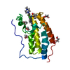
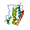
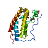
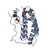
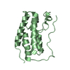
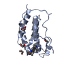
| Entry | Database: PDB / ID: 6sah | ||||||
|---|---|---|---|---|---|---|---|
| Title | Crystal Structure of BRD4(1) bound to inhibitor BUX5 (11) | ||||||
 Components Components | Bromodomain-containing protein 4 | ||||||
 Keywords Keywords | TRANSCRIPTION / BRD4 / BRD4(1) / INHIBITOR / BROMODOMAIN / EPIGENETIC READER PROTEIN / ACETYLATED / LYSINE / HISTONE TAIL / PROTEIN BINDING-INHIBITOR COMPLEX / BUX5 / fragment | ||||||
| Function / homology |  Function and homology information Function and homology informationhistone H4K8ac reader activity / RNA polymerase II C-terminal domain binding / histone H3K27ac reader activity / P-TEFb complex binding / histone H3K9ac reader activity / negative regulation of DNA damage checkpoint / histone H4 reader activity / histone H4K5ac reader activity / histone H4K12ac reader activity / host-mediated suppression of viral transcription ...histone H4K8ac reader activity / RNA polymerase II C-terminal domain binding / histone H3K27ac reader activity / P-TEFb complex binding / histone H3K9ac reader activity / negative regulation of DNA damage checkpoint / histone H4 reader activity / histone H4K5ac reader activity / histone H4K12ac reader activity / host-mediated suppression of viral transcription / histone H4K16ac reader activity / positive regulation of G2/M transition of mitotic cell cycle / positive regulation of T-helper 17 cell lineage commitment / RNA polymerase II CTD heptapeptide repeat kinase activity / condensed nuclear chromosome / transcription coregulator activity / positive regulation of transcription elongation by RNA polymerase II / p53 binding / chromosome / regulation of inflammatory response / histone binding / Potential therapeutics for SARS / positive regulation of canonical NF-kappaB signal transduction / transcription coactivator activity / transcription cis-regulatory region binding / chromatin remodeling / protein serine/threonine kinase activity / chromatin binding / regulation of transcription by RNA polymerase II / DNA damage response / positive regulation of DNA-templated transcription / chromatin / enzyme binding / positive regulation of transcription by RNA polymerase II / nucleoplasm / nucleus Similarity search - Function | ||||||
| Biological species |  Homo sapiens (human) Homo sapiens (human) | ||||||
| Method |  X-RAY DIFFRACTION / SYNCHROTRON / MOLECULAR REPLACEMENT / molecular replacement / Resolution: 1.5 Å X-RAY DIFFRACTION / SYNCHROTRON / MOLECULAR REPLACEMENT / molecular replacement / Resolution: 1.5 Å | ||||||
 Authors Authors | Huegle, M. | ||||||
| Funding support |  Germany, 1items Germany, 1items
| ||||||
 Citation Citation | Journal: J.Med.Chem. / Year: 2020 Title: 4-Acyl Pyrroles as Dual BET-BRD7/9 Bromodomain Inhibitors Address BETi Insensitive Human Cancer Cell Lines. Authors: Hugle, M. / Regenass, P. / Warstat, R. / Hau, M. / Schmidtkunz, K. / Lucas, X. / Wohlwend, D. / Einsle, O. / Jung, M. / Breit, B. / Gunther, S. | ||||||
| History |
|
- Structure visualization
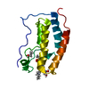
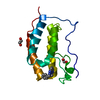
| Structure viewer | Molecule: MolmilJmol/JSmol |
|---|
- Downloads & links
-Download
| PDBx/mmCIF format | 6sah.cif.gz | 47.8 KB | Display | PDBx/mmCIF format |
|---|---|---|---|---|
| PDB format | pdb6sah.ent.gz | 31 KB | Display | PDB format |
| PDBx/mmJSON format | 6sah.json.gz | Tree view | PDBx/mmJSON format | |
| Others |  Other downloads Other downloads |
-Validation report
| Arichive directory | https://data.pdbj.org/pub/pdb/validation_reports/sa/6sahftp://data.pdbj.org/pub/pdb/validation_reports/sa/6sah | HTTPS FTP |
|---|
-Related structure data
| Related structure data | 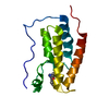 6rwjC 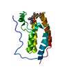 6s4bC 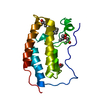 6s6kC 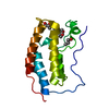 6sa2C 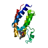 6sa3C 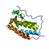 6sajC 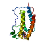 6sb8C 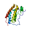 4lywS S: Starting model for refinement C: citing same article ( |
|---|---|
| Similar structure data |


-Links
 PDBj
PDBj- Assembly
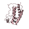
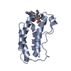
| Deposited unit | 
| ||||||||
|---|---|---|---|---|---|---|---|---|---|
| 1 |
| ||||||||
| Unit cell |
|
-Components
| #1: Protein | Mass: 15099.380 Da / Num. of mol.: 1 Source method: isolated from a genetically manipulated source Source: (gene. exp.) Homo sapiens (human) / Gene: BRD4, HUNK1 / Production host:  |
|---|---|
| #2: Chemical | ChemComp-L2W / ~{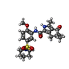  Mass: 459.558 Da / Num. of mol.: 1 / Source method: obtained synthetically / Formula: C23H29N3O5S / Feature type: SUBJECT OF INVESTIGATION Mass: 459.558 Da / Num. of mol.: 1 / Source method: obtained synthetically / Formula: C23H29N3O5S / Feature type: SUBJECT OF INVESTIGATION |
| #3: Water | ChemComp-HOH /  Mass: 18.015 Da / Num. of mol.: 188 / Source method: isolated from a natural source / Formula: H2O Mass: 18.015 Da / Num. of mol.: 188 / Source method: isolated from a natural source / Formula: H2O |
| Has ligand of interest | Y |
-Experimental details
-Experiment
| Experiment | Method: X-RAY DIFFRACTION / Number of used crystals: 1 |
|---|
- Sample preparation
| Crystal | Density Matthews: 1.79 Å3/Da / Density % sol: 31.41 % |
|---|---|
| Crystal grow | Temperature: 293 K / Method: vapor diffusion, sitting drop / pH: 5.5 Details: Index D6, 0.1 M BIS-TRIS pH 5.5, 25% w/v Polyethylene glycol 3,350 |
-Data collection
| Diffraction | Mean temperature: 100 K / Serial crystal experiment: N | ||||||||||||||||||||||||||||||
|---|---|---|---|---|---|---|---|---|---|---|---|---|---|---|---|---|---|---|---|---|---|---|---|---|---|---|---|---|---|---|---|
| Diffraction source | Source: SYNCHROTRON / Site: SLS  / Beamline: X06SA / Wavelength: 0.99998 Å / Beamline: X06SA / Wavelength: 0.99998 Å | ||||||||||||||||||||||||||||||
| Detector | Type: DECTRIS EIGER X 16M / Detector: PIXEL / Date: Sep 16, 2018 | ||||||||||||||||||||||||||||||
| Radiation | Protocol: SINGLE WAVELENGTH / Monochromatic (M) / Laue (L): M / Scattering type: x-ray | ||||||||||||||||||||||||||||||
| Radiation wavelength | Wavelength: 0.99998 Å / Relative weight: 1 | ||||||||||||||||||||||||||||||
| Reflection | Resolution: 1.5→46.22 Å / Num. obs: 17986 / % possible obs: 99.9 % / Redundancy: 12.8 % / CC1/2: 0.999 / Rmerge(I) obs: 0.048 / Rpim(I) all: 0.014 / Rrim(I) all: 0.05 / Net I/σ(I): 30.4 / Num. measured all: 230945 / Scaling rejects: 5 | ||||||||||||||||||||||||||||||
| Reflection shell | Diffraction-ID: 1
|
-Phasing
| Phasing | Method: molecular replacement | |||||||||
|---|---|---|---|---|---|---|---|---|---|---|
| Phasing MR |
|
- Processing
| Software |
| |||||||||||||||||||||||||||||||||||
|---|---|---|---|---|---|---|---|---|---|---|---|---|---|---|---|---|---|---|---|---|---|---|---|---|---|---|---|---|---|---|---|---|---|---|---|---|
| Refinement | Method to determine structure: MOLECULAR REPLACEMENT Starting model: 4LYW Resolution: 1.5→35.907 Å / SU ML: 0.12 / Cross valid method: THROUGHOUT / σ(F): 1.38 / Phase error: 17.3
| |||||||||||||||||||||||||||||||||||
| Solvent computation | Shrinkage radii: 0.9 Å / VDW probe radii: 1.11 Å | |||||||||||||||||||||||||||||||||||
| Displacement parameters | Biso max: 79.83 Å2 / Biso mean: 19.1059 Å2 / Biso min: 8.46 Å2 | |||||||||||||||||||||||||||||||||||
| Refinement step | Cycle: final / Resolution: 1.5→35.907 Å
| |||||||||||||||||||||||||||||||||||
| LS refinement shell | Refine-ID: X-RAY DIFFRACTION / Rfactor Rfree error: 0 / % reflection obs: 100 %
|
