[English] 日本語
Yorodumi
- PDB-6fm4: The crystal structure of S. aureus Gyrase complex with ID-130 and DNA -

+
Open data


ID or keywords:

Loading...

-
Basic information

Entry
Database: PDB / ID: 6fm4
TitleThe crystal structure of S. aureus Gyrase complex with ID-130 and DNA
Components
  • DNA (5'-5UA*D(P*GP*CP*CP*GP*TP*AP*GP*GP*GP*CP*CP*CP*TP*AP*CP*GP*GP*C)-3')
  • DNA (5'-5UA*D(P*GP*CP*CP*GP*TP*AP*GP*GP*GP*CP*CP*CP*TP*AP*CP*GP*GP*CP*T)-3')
  • DNA gyrase subunit B,DNA gyrase subunit B,DNA gyrase subunit A
KeywordsISOMERASE / NBTIs / BACTERIAL TOPOISOMERASE / GYRASE
Function / homology
Function and homology information


DNA negative supercoiling activity / DNA topoisomerase type II (double strand cut, ATP-hydrolyzing) complex / DNA topoisomerase type II (double strand cut, ATP-hydrolyzing) activity / DNA topoisomerase (ATP-hydrolysing) / DNA topological change / DNA-templated DNA replication / chromosome / response to antibiotic / DNA binding / ATP binding ...DNA negative supercoiling activity / DNA topoisomerase type II (double strand cut, ATP-hydrolyzing) complex / DNA topoisomerase type II (double strand cut, ATP-hydrolyzing) activity / DNA topoisomerase (ATP-hydrolysing) / DNA topological change / DNA-templated DNA replication / chromosome / response to antibiotic / DNA binding / ATP binding / metal ion binding / cytoplasm
Similarity search - Function
Rossmann fold - #670 / DNA gyrase, subunit A / DNA gyrase/topoisomerase IV, subunit A, C-terminal repeat / DNA gyrase/topoisomerase IV, subunit A, C-terminal / DNA gyrase C-terminal domain, beta-propeller / Topoisomerase (Topo) IIA-type catalytic domain profile. / DNA gyrase subunit B, TOPRIM domain / DNA gyrase, subunit B / DNA topoisomerase, type IIA, subunit B / DNA topoisomerase, type IIA, alpha-helical domain superfamily ...Rossmann fold - #670 / DNA gyrase, subunit A / DNA gyrase/topoisomerase IV, subunit A, C-terminal repeat / DNA gyrase/topoisomerase IV, subunit A, C-terminal / DNA gyrase C-terminal domain, beta-propeller / Topoisomerase (Topo) IIA-type catalytic domain profile. / DNA gyrase subunit B, TOPRIM domain / DNA gyrase, subunit B / DNA topoisomerase, type IIA, subunit B / DNA topoisomerase, type IIA, alpha-helical domain superfamily / DNA topoisomerase, type IIA, domain A / DNA topoisomerase, type IIA, domain A, alpha-beta / DNA gyrase/topoisomerase IV, subunit A / DNA Topoisomerase IV / DNA gyrase B subunit, C-terminal / DNA gyrase B subunit, carboxyl terminus / DNA topoisomerase, type IIA, subunit B, domain 2 / DNA gyrase B / DNA topoisomerase, type IIA / DNA topoisomerase, type IIA, conserved site / DNA topoisomerase II signature. / TopoisomeraseII / DNA topoisomerase, type IIA, subunit B, C-terminal / Toprim domain / DNA topoisomerase, type IIA-like domain superfamily / Toprim domain profile. / TOPRIM domain / Histidine kinase-, DNA gyrase B-, and HSP90-like ATPase / Histidine kinase-like ATPases / Histidine kinase/HSP90-like ATPase / Histidine kinase/HSP90-like ATPase superfamily / Ribosomal protein S5 domain 2-type fold, subgroup / Ribosomal protein S5 domain 2-type fold / Rossmann fold / 3-Layer(aba) Sandwich / Alpha Beta
Similarity search - Domain/homology
5'-O-CARBOXY-2'-DEOXYADENOSINE / Chem-DU5 / : / DNA / DNA (> 10) / DNA gyrase subunit B / DNA gyrase subunit A
Similarity search - Component
Biological speciesStaphylococcus aureus (bacteria)
synthetic construct (others)
MethodX-RAY DIFFRACTION / SYNCHROTRON / MOLECULAR REPLACEMENT / Resolution: 2.7 Å
AuthorsOmbrato, R. / Garofalo, B. / Mangano, G. / Mancini, F.
CitationJournal: J.Med.Chem. / Year: 2019
Title: Virtual Screening Approach and Investigation of Structure-Activity Relationships To Discover Novel Bacterial Topoisomerase Inhibitors Targeting Gram-Positive and Gram-Negative Pathogens.
Authors: Magaro, G. / Prati, F. / Garofalo, B. / Corso, G. / Furlotti, G. / Apicella, C. / Mangano, G. / D'Atanasio, N. / Robinson, D. / Di Giorgio, F.P. / Ombrato, R.
History
DepositionJan 30, 2018Deposition site: PDBE / Processing site: PDBE
Revision 1.0Jul 10, 2019Provider: repository / Type: Initial release
Revision 1.1Jul 17, 2019Group: Data collection / Database references / Category: citation / citation_author
Item: _citation.country / _citation.journal_abbrev ..._citation.country / _citation.journal_abbrev / _citation.journal_id_ASTM / _citation.journal_id_CSD / _citation.journal_id_ISSN / _citation.pdbx_database_id_DOI / _citation.pdbx_database_id_PubMed / _citation.title / _citation.year
Revision 1.2Aug 7, 2019Group: Data collection / Database references / Category: citation / citation_author
Item: _citation.title / _citation_author.identifier_ORCID / _citation_author.name
Revision 1.3Sep 4, 2019Group: Data collection / Database references / Category: citation / citation_author
Item: _citation.journal_volume / _citation.page_first ..._citation.journal_volume / _citation.page_first / _citation.page_last / _citation_author.identifier_ORCID
Revision 1.4Jan 17, 2024Group: Advisory / Data collection ...Advisory / Data collection / Database references / Derived calculations / Refinement description
Category: chem_comp_atom / chem_comp_bond ...chem_comp_atom / chem_comp_bond / database_2 / pdbx_initial_refinement_model / pdbx_unobs_or_zero_occ_residues / struct_conn / struct_conn_type
Item: _database_2.pdbx_DOI / _database_2.pdbx_database_accession ..._database_2.pdbx_DOI / _database_2.pdbx_database_accession / _struct_conn.conn_type_id / _struct_conn.id / _struct_conn.pdbx_dist_value / _struct_conn.pdbx_leaving_atom_flag / _struct_conn.ptnr1_auth_asym_id / _struct_conn.ptnr1_auth_comp_id / _struct_conn.ptnr1_auth_seq_id / _struct_conn.ptnr1_label_asym_id / _struct_conn.ptnr1_label_atom_id / _struct_conn.ptnr1_label_comp_id / _struct_conn.ptnr1_label_seq_id / _struct_conn.ptnr2_auth_asym_id / _struct_conn.ptnr2_auth_comp_id / _struct_conn.ptnr2_auth_seq_id / _struct_conn.ptnr2_label_asym_id / _struct_conn.ptnr2_label_atom_id / _struct_conn.ptnr2_label_comp_id / _struct_conn_type.id

-
Structure visualization

Structure viewerMolecule:
MolmilJmol/JSmol

Downloads & links

-
Assembly

Deposited unit
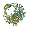
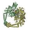
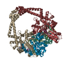
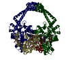
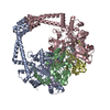
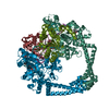
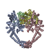
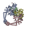
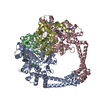
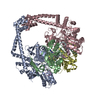
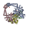
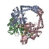
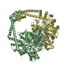
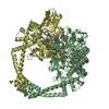
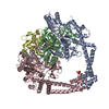
B: DNA gyrase subunit B,DNA gyrase subunit B,DNA gyrase subunit A
D: DNA gyrase subunit B,DNA gyrase subunit B,DNA gyrase subunit A
E: DNA (5'-5UA*D(P*GP*CP*CP*GP*TP*AP*GP*GP*GP*CP*CP*CP*TP*AP*CP*GP*GP*CP*T)-3')
F: DNA (5'-5UA*D(P*GP*CP*CP*GP*TP*AP*GP*GP*GP*CP*CP*CP*TP*AP*CP*GP*GP*C)-3')
hetero molecules


Theoretical massNumber of molelcules
Total (without water)168,68910
Polymers167,5594
Non-polymers1,1306
Water1,11762
1


  • Idetical with deposited unit
  • defined by author&software
TypeNameSymmetry operationNumber
identity operation1_555x,y,z1
Buried area15820 Å2
ΔGint-87 kcal/mol
Surface area56920 Å2
MethodPISA
Unit cell
Length a, b, c (Å)92.951, 92.951, 407.030
Angle α, β, γ (deg.)90.000, 90.000, 120.000
Int Tables number169
Space group name H-MP61

-
Components

-
Protein , 1 types, 2 molecules BD

#1: Protein DNA gyrase subunit B,DNA gyrase subunit B,DNA gyrase subunit A


Mass: 78109.000 Da / Num. of mol.: 2 / Mutation: Y1123F
Source method: isolated from a genetically manipulated source
Details: C-TERMINUS GYRB (640) FUSED TO N-TERMINUS GYRA (1010). DOMAIN 544-579 DELETED AND REPLACED WITH TWO RESIDUES TG.
Source: (gene. exp.) Staphylococcus aureus (strain N315) (bacteria)
Gene: gyrB, SA0005, gyrA, SA0006 / Production host: Escherichia coli (E. coli) / References: UniProt: P66937, UniProt: Q99XG5, EC: 5.99.1.3

-
DNA chain , 2 types, 2 molecules EF

#2: DNA chain DNA (5'-5UA*D(P*GP*CP*CP*GP*TP*AP*GP*GP*GP*CP*CP*CP*TP*AP*CP*GP*GP*CP*T)-3')


Mass: 5822.748 Da / Num. of mol.: 1 / Source method: obtained synthetically / Source: (synth.) synthetic construct (others)
#3: DNA chain DNA (5'-5UA*D(P*GP*CP*CP*GP*TP*AP*GP*GP*GP*CP*CP*CP*TP*AP*CP*GP*GP*C)-3')


Mass: 5518.555 Da / Num. of mol.: 1 / Source method: obtained synthetically / Source: (synth.) synthetic construct (others)

-
Non-polymers , 4 types, 68 molecules

#4: Chemical ChemComp-MN / MANGANESE (II) ION


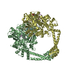
Mass: 54.938 Da / Num. of mol.: 3 / Source method: obtained synthetically / Formula: Mn
#5: Chemical ChemComp-5UA / 5'-O-CARBOXY-2'-DEOXYADENOSINE


Type: DNA linking / Mass: 295.251 Da / Num. of mol.: 2 / Source method: obtained synthetically / Formula: C11H13N5O5
#6: Chemical ChemComp-DU5 / ~{N}-[3-(4-isoquinolin-1-ylpiperazin-1-yl)propyl]benzamide


Mass: 374.479 Da / Num. of mol.: 1 / Source method: obtained synthetically / Formula: C23H26N4O / Feature type: SUBJECT OF INVESTIGATION
#7: Water ChemComp-HOH / water


Mass: 18.015 Da / Num. of mol.: 62 / Source method: isolated from a natural source / Formula: H2O

-
Experimental details

-
Experiment

ExperimentMethod: X-RAY DIFFRACTION / Number of used crystals: 1

-
Sample preparation

CrystalDensity Matthews: 3.11 Å3/Da / Density % sol: 60.44 %
Crystal growTemperature: 295.15 K / Method: vapor diffusion, hanging drop / pH: 6.4 / Details: 18% PEG 5000 MME, 0.1 M BISTRIS PH 6.5

-
Data collection

DiffractionMean temperature: 100 K
Diffraction sourceSource: SYNCHROTRON / Site: SSRF / Beamline: BL17U1 / Wavelength: 0.9 Å
DetectorType: ADSC QUANTUM 315r / Detector: CCD / Date: Sep 25, 2013
RadiationProtocol: SINGLE WAVELENGTH / Monochromatic (M) / Laue (L): M / Scattering type: x-ray
Radiation wavelengthWavelength: 0.9 Å / Relative weight: 1
ReflectionResolution: 2.7→43.05 Å / Num. obs: 51255 / % possible obs: 99.7 % / Redundancy: 7.5 % / Rmerge(I) obs: 0.084 / Net I/σ(I): 30.68
Reflection shellResolution: 2.7→2.77 Å

-
Processing

Software
NameVersionClassification
HKL-2000data scaling
REFMAC5.7.0032refinement
PDB_EXTRACT3.24data extraction
RefinementMethod to determine structure: MOLECULAR REPLACEMENT
Starting model: 2XCS
Resolution: 2.7→43.05 Å / Cor.coef. Fo:Fc: 0.885 / Cor.coef. Fo:Fc free: 0.794 / SU B: 15.544 / SU ML: 0.324 / Cross valid method: THROUGHOUT / ESU R: 0.911 / ESU R Free: 0.391
Details: HYDROGENS HAVE BEEN ADDED IN THE RIDING POSITIONS U VALUES: REFINED INDIVIDUALLY
RfactorNum. reflection% reflectionSelection details
Rfree0.2999 2750 5.1 %RANDOM
Rwork0.2245 ---
obs0.2284 51255 99.7 %-
Solvent computationIon probe radii: 0.8 Å / Shrinkage radii: 0.8 Å / VDW probe radii: 1.2 Å
Displacement parametersBiso max: 102.83 Å2 / Biso mean: 39.691 Å2 / Biso min: 16.2 Å2
Baniso -1Baniso -2Baniso -3
1--0.8 Å2-0.8 Å2-0 Å2
2---0.8 Å2-0 Å2
3---2.58 Å2
Refinement stepCycle: final / Resolution: 2.7→43.05 Å
ProteinNucleic acidLigandSolventTotal
Num. atoms10606 758 56 62 11482
Biso mean--59.05 29.71 -
Num. residues----1379
Refine LS restraints
Refine-IDTypeDev idealDev ideal targetNumber
X-RAY DIFFRACTIONr_bond_refined_d0.0130.01911668
X-RAY DIFFRACTIONr_bond_other_d0.0020.0210993
X-RAY DIFFRACTIONr_angle_refined_deg1.6741.91515885
X-RAY DIFFRACTIONr_angle_other_deg0.9713.00225231
X-RAY DIFFRACTIONr_dihedral_angle_1_deg6.84451338
X-RAY DIFFRACTIONr_dihedral_angle_2_deg35.12923.67534
X-RAY DIFFRACTIONr_dihedral_angle_3_deg15.439152001
X-RAY DIFFRACTIONr_dihedral_angle_4_deg16.9815118
X-RAY DIFFRACTIONr_chiral_restr0.0820.21748
X-RAY DIFFRACTIONr_gen_planes_refined0.0070.0212718
X-RAY DIFFRACTIONr_gen_planes_other0.0010.022656
LS refinement shellResolution: 2.702→2.772 Å / Rfactor Rfree error: 0 / Total num. of bins used: 20
RfactorNum. reflection% reflection
Rfree0.34 201 -
Rwork0.252 3767 -
all-3968 -
obs--99.25 %

+
About Yorodumi

-
News

-
Feb 9, 2022. New format data for meta-information of EMDB entries

New format data for meta-information of EMDB entries

  • Version 3 of the EMDB header file is now the official format.
  • The previous official version 1.9 will be removed from the archive.

Related info.:EMDB header

External links:wwPDB to switch to version 3 of the EMDB data model

-
Aug 12, 2020. Covid-19 info

Covid-19 info

URL: https://pdbjlvh1.pdbj.org/emnavi/covid19.php

New page: Covid-19 featured information page in EM Navigator.

Related info.:Covid-19 info / Mar 5, 2020. Novel coronavirus structure data

+
Mar 5, 2020. Novel coronavirus structure data

Novel coronavirus structure data

Related info.:Yorodumi Speices / Aug 12, 2020. Covid-19 info

External links:COVID-19 featured content - PDBj / Molecule of the Month (242):Coronavirus Proteases

+
Jan 31, 2019. EMDB accession codes are about to change! (news from PDBe EMDB page)

EMDB accession codes are about to change! (news from PDBe EMDB page)

  • The allocation of 4 digits for EMDB accession codes will soon come to an end. Whilst these codes will remain in use, new EMDB accession codes will include an additional digit and will expand incrementally as the available range of codes is exhausted. The current 4-digit format prefixed with “EMD-” (i.e. EMD-XXXX) will advance to a 5-digit format (i.e. EMD-XXXXX), and so on. It is currently estimated that the 4-digit codes will be depleted around Spring 2019, at which point the 5-digit format will come into force.
  • The EM Navigator/Yorodumi systems omit the EMD- prefix.

Related info.:Q: What is EMD? / ID/Accession-code notation in Yorodumi/EM Navigator

External links:EMDB Accession Codes are Changing Soon! / Contact to PDBj

+
Jul 12, 2017. Major update of PDB

Major update of PDB

  • wwPDB released updated PDB data conforming to the new PDBx/mmCIF dictionary.
  • This is a major update changing the version number from 4 to 5, and with Remediation, in which all the entries are updated.
  • In this update, many items about electron microscopy experimental information are reorganized (e.g. em_software).
  • Now, EM Navigator and Yorodumi are based on the updated data.

External links:wwPDB Remediation / Enriched Model Files Conforming to OneDep Data Standards Now Available in the PDB FTP Archive

-
Yorodumi

Thousand views of thousand structures

  • Yorodumi is a browser for structure data from EMDB, PDB, SASBDB, etc.
  • This page is also the successor to EM Navigator detail page, and also detail information page/front-end page for Omokage search.
  • The word "yorodu" (or yorozu) is an old Japanese word meaning "ten thousand". "mi" (miru) is to see.

Related info.:EMDB / PDB / SASBDB / Comparison of 3 databanks / Yorodumi Search / Aug 31, 2016. New EM Navigator & Yorodumi / Yorodumi Papers / Jmol/JSmol / Function and homology information / Changes in new EM Navigator and Yorodumi

Read more