 Movie
Movie Controller


+ Open data

- Basic information
| Entry | Database: PDB / ID: 6con | ||||||
|---|---|---|---|---|---|---|---|
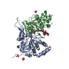
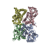
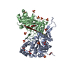
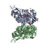
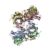
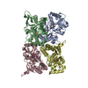
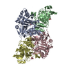
| Title | Crystal structure of Mycobacterium tuberculosis IpdAB | ||||||
 Components Components |
| ||||||
 Keywords Keywords | HYDROLASE / Cholesterol / Ring cleaving / virulence factor | ||||||
| Function / homology |  Function and homology information Function and homology information3-oxoadipate CoA-transferase / 3-oxoadipate CoA-transferase activity / glutaconate CoA-transferase / glutaconate CoA-transferase activity / Lyases; Carbon-carbon lyases; Other carbon-carbon lyases / Transferases; Transferring sulfur-containing groups; CoA-transferases / CoA-transferase activity / biological process involved in interaction with host / cholesterol catabolic process / lyase activity Similarity search - Function | ||||||
| Biological species |   Mycobacterium tuberculosis (bacteria) Mycobacterium tuberculosis (bacteria) | ||||||
| Method |  X-RAY DIFFRACTION / SYNCHROTRON / MOLECULAR REPLACEMENT / Resolution: 2.1 Å X-RAY DIFFRACTION / SYNCHROTRON / MOLECULAR REPLACEMENT / Resolution: 2.1 Å | ||||||
 Authors Authors | Crowe, A.M. / Workman, S.D. / Watanabe, N. / Worrall, L.J. / Strynadka, N.C.J. / Eltis, L.D. | ||||||
 Citation Citation | Journal: Proc. Natl. Acad. Sci. U.S.A. / Year: 2018 Title: IpdAB, a virulence factor inMycobacterium tuberculosis, is a cholesterol ring-cleaving hydrolase. Authors: Crowe, A.M. / Workman, S.D. / Watanabe, N. / Worrall, L.J. / Strynadka, N.C.J. / Eltis, L.D. | ||||||
| History |
|
- Structure visualization
| Structure viewer | Molecule: MolmilJmol/JSmol |
|---|
- Downloads & links
-Download
| PDBx/mmCIF format | 6con.cif.gz | 430.1 KB | Display | PDBx/mmCIF format |
|---|---|---|---|---|
| PDB format | pdb6con.ent.gz | 351.1 KB | Display | PDB format |
| PDBx/mmJSON format | 6con.json.gz | Tree view | PDBx/mmJSON format | |
| Others |  Other downloads Other downloads |
-Validation report
| Arichive directory | https://data.pdbj.org/pub/pdb/validation_reports/co/6conftp://data.pdbj.org/pub/pdb/validation_reports/co/6con | HTTPS FTP |
|---|
-Related structure data
| Related structure data |  6co6SC  6co9C  6cojC S: Starting model for refinement C: citing same article ( |
|---|---|
| Similar structure data |


-Links
 PDBj
PDBj- Assembly
| Deposited unit | 
| ||||||||
|---|---|---|---|---|---|---|---|---|---|
| 1 | 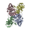
| ||||||||
| 2 | 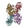
| ||||||||
| Unit cell |
|
-Components
| #1: Protein | Mass: 33325.703 Da / Num. of mol.: 4 Source method: isolated from a genetically manipulated source Source: (gene. exp.) Mycobacterium tuberculosis (bacteria)Gene: gctA, CRX58_09925, ERS007663_04080, ERS007665_02371, ERS023446_02783, ERS027646_00148, ERS027654_03521, ERS027656_00696, ERS124361_01188, SAMEA2682864_02618, SAMEA2683035_00650 Production host: Rhodococcus jostii RHA1 (bacteria)References: UniProt: A0A045J8X5, UniProt: P9WPW1*PLUS, Transferases; Transferring sulfur-containing groups; CoA-transferases, glutaconate CoA-transferase #2: Protein | Mass: 27426.020 Da / Num. of mol.: 4 Source method: isolated from a genetically manipulated source Source: (gene. exp.) Mycobacterium tuberculosis (bacteria)Gene: catJ, CRX58_09930, ERS007657_01926, ERS007661_02237, ERS007665_02370, ERS007670_03185, ERS007672_03945, ERS007679_04090, ERS007681_03977, ERS007688_03438, ERS007703_04697, ERS007722_01813, ...Gene: catJ, CRX58_09930, ERS007657_01926, ERS007661_02237, ERS007665_02370, ERS007670_03185, ERS007672_03945, ERS007679_04090, ERS007681_03977, ERS007688_03438, ERS007703_04697, ERS007722_01813, ERS007741_03174, ERS023446_02782, ERS024213_01521, ERS024276_02483, ERS027644_04603, ERS027646_00147, ERS027656_00695, ERS027659_03102, ERS027661_03605, ERS027666_03707, ERS124361_01187, SAMEA2682864_02619, SAMEA2683035_00651 Production host: Rhodococcus jostii RHA1 (bacteria)References: UniProt: A0A045H5Z8, UniProt: P9WPV9*PLUS, Transferases; Transferring sulfur-containing groups; CoA-transferases, 3-oxoadipate CoA-transferase #3: Water | ChemComp-HOH / |  Mass: 18.015 Da / Num. of mol.: 981 / Source method: isolated from a natural source / Formula: H2O Mass: 18.015 Da / Num. of mol.: 981 / Source method: isolated from a natural source / Formula: H2O |
|---|
-Experimental details
-Experiment
| Experiment | Method: X-RAY DIFFRACTION / Number of used crystals: 1 |
|---|
- Sample preparation
| Crystal | Density Matthews: 2.25 Å3/Da / Density % sol: 45.37 % |
|---|---|
| Crystal grow | Temperature: 293 K / Method: vapor diffusion, sitting drop / Details: 25% PEG 3350, 0.5 M magnesium formate |
-Data collection
| Diffraction | Mean temperature: 100 K | ||||||||||||||||||||||||||||||
|---|---|---|---|---|---|---|---|---|---|---|---|---|---|---|---|---|---|---|---|---|---|---|---|---|---|---|---|---|---|---|---|
| Diffraction source | Source: SYNCHROTRON / Site: CLSI  / Beamline: 08B1-1 / Wavelength: 0.97777 Å / Beamline: 08B1-1 / Wavelength: 0.97777 Å | ||||||||||||||||||||||||||||||
| Detector | Type: RAYONIX MX300HE / Detector: CCD / Date: Aug 7, 2013 | ||||||||||||||||||||||||||||||
| Radiation | Protocol: SINGLE WAVELENGTH / Monochromatic (M) / Laue (L): M / Scattering type: x-ray | ||||||||||||||||||||||||||||||
| Radiation wavelength | Wavelength: 0.97777 Å / Relative weight: 1 | ||||||||||||||||||||||||||||||
| Reflection | Resolution: 2.1→88.86 Å / Num. obs: 119998 / % possible obs: 98.7 % / Redundancy: 3.8 % / CC1/2: 0.995 / Rmerge(I) obs: 0.07 / Rpim(I) all: 0.041 / Rrim(I) all: 0.081 / Net I/σ(I): 12.2 / Num. measured all: 456667 / Scaling rejects: 38 | ||||||||||||||||||||||||||||||
| Reflection shell | Diffraction-ID: 1
|
- Processing
| Software |
| ||||||||||||||||||||||||
|---|---|---|---|---|---|---|---|---|---|---|---|---|---|---|---|---|---|---|---|---|---|---|---|---|---|
| Refinement | Method to determine structure: MOLECULAR REPLACEMENT Starting model: 6CO6 Resolution: 2.1→88.86 Å / Cor.coef. Fo:Fc: 0.869 / Cor.coef. Fo:Fc free: 0.854 / WRfactor Rfree: 0.2764 / WRfactor Rwork: 0.2523 / FOM work R set: 0.8159 / SU B: 0.002 / SU ML: 0 / SU R Cruickshank DPI: 0.2117 / SU Rfree: 0.2313 / Cross valid method: THROUGHOUT / σ(F): 0 / ESU R: 0.212 / ESU R Free: 0.231 / Stereochemistry target values: MAXIMUM LIKELIHOOD Details: HYDROGENS HAVE BEEN ADDED IN THE RIDING POSITIONS U VALUES : REFINED INDIVIDUALLY
| ||||||||||||||||||||||||
| Solvent computation | Ion probe radii: 0.8 Å / Shrinkage radii: 0.8 Å / VDW probe radii: 1.2 Å / Solvent model: MASK | ||||||||||||||||||||||||
| Displacement parameters | Biso max: 100.33 Å2 / Biso mean: 25.566 Å2 / Biso min: 9.71 Å2
| ||||||||||||||||||||||||
| Refinement step | Cycle: final / Resolution: 2.1→88.86 Å
| ||||||||||||||||||||||||
| LS refinement shell | Resolution: 2.1→2.155 Å / Rfactor Rfree error: 0 / Total num. of bins used: 20
|
