 Movie
Movie Controller


+ Open data

- Basic information
| Entry | Database: PDB / ID: 5mzw | ||||||
|---|---|---|---|---|---|---|---|
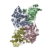
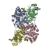
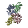
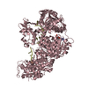
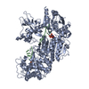
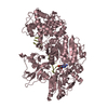
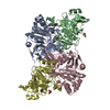
| Title | Crystal structure of the decarboxylase AibA/AibB | ||||||
 Components Components |
| ||||||
 Keywords Keywords | LYASE / decarboxylase / CoA transferase like fold | ||||||
| Function / homology | CoA-transferase activity / Coenzyme A transferase family I / Coenzyme A transferase / Coenzyme A transferase / NagB/RpiA transferase-like / Glutaconate CoA-transferase family, subunit B / Glutaconate CoA-transferase family, subunit A Function and homology information Function and homology information | ||||||
| Biological species |  Myxococcus xanthus (bacteria) Myxococcus xanthus (bacteria) | ||||||
| Method |  X-RAY DIFFRACTION / SYNCHROTRON / SAD / Resolution: 1.52 Å X-RAY DIFFRACTION / SYNCHROTRON / SAD / Resolution: 1.52 Å | ||||||
 Authors Authors | Bock, T. / Luxenburger, E. / Hoffmann, J. / Schuetza, V. / Feiler, C. / Mueller, R. / Blankenfeldt, W. | ||||||
 Citation Citation | Journal: Angew. Chem. Int. Ed. Engl. / Year: 2017 Title: AibA/AibB Induces an Intramolecular Decarboxylation in Isovalerate Biosynthesis by Myxococcus xanthus. Authors: Bock, T. / Luxenburger, E. / Hoffmann, J. / Schutza, V. / Feiler, C. / Muller, R. / Blankenfeldt, W. | ||||||
| History |
|
- Structure visualization
| Structure viewer | Molecule: MolmilJmol/JSmol |
|---|
- Downloads & links
-Download
| PDBx/mmCIF format | 5mzw.cif.gz | 384.4 KB | Display | PDBx/mmCIF format |
|---|---|---|---|---|
| PDB format | pdb5mzw.ent.gz | 313 KB | Display | PDB format |
| PDBx/mmJSON format | 5mzw.json.gz | Tree view | PDBx/mmJSON format | |
| Others |  Other downloads Other downloads |
-Validation report
| Arichive directory | https://data.pdbj.org/pub/pdb/validation_reports/mz/5mzwftp://data.pdbj.org/pub/pdb/validation_reports/mz/5mzw | HTTPS FTP |
|---|
-Related structure data
| Related structure data | 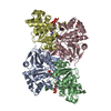 5mzxC  5mzyC  5mzzC 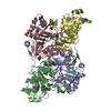 5n00C  5n01C  5n02C  5n03C C: citing same article ( |
|---|---|
| Similar structure data |


-Links
 PDBj
PDBj- Assembly
| Deposited unit | 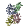
| ||||||||
|---|---|---|---|---|---|---|---|---|---|
| 1 |
| ||||||||
| Unit cell |
|
-Components
| #1: Protein | Mass: 28367.838 Da / Num. of mol.: 2 / Mutation: K191A Source method: isolated from a genetically manipulated source Source: (gene. exp.) Myxococcus xanthus (strain DK 1622) (bacteria)Strain: DK 1622 / Gene: MXAN_4264 / Production host: #2: Protein | Mass: 26280.871 Da / Num. of mol.: 2 / Mutation: E200A, E201A Source method: isolated from a genetically manipulated source Source: (gene. exp.) Myxococcus xanthus (strain DK 1622) (bacteria)Strain: DK 1622 / Gene: MXAN_4265 / Production host: #3: Chemical |   Mass: 92.094 Da / Num. of mol.: 3 / Source method: obtained synthetically / Formula: C3H8O3 Mass: 92.094 Da / Num. of mol.: 3 / Source method: obtained synthetically / Formula: C3H8O3#4: Water | ChemComp-HOH / |  Mass: 18.015 Da / Num. of mol.: 1226 / Source method: isolated from a natural source / Formula: H2O Mass: 18.015 Da / Num. of mol.: 1226 / Source method: isolated from a natural source / Formula: H2O |
|---|
-Experimental details
-Experiment
| Experiment | Method: X-RAY DIFFRACTION / Number of used crystals: 1 |
|---|
- Sample preparation
| Crystal | Density Matthews: 2.5 Å3/Da / Density % sol: 50.8 % |
|---|---|
| Crystal grow | Temperature: 293 K / Method: vapor diffusion, sitting drop / Details: 0.1 M MES pH 5.0, 12 % PEG6000 |
-Data collection
| Diffraction | Mean temperature: 100 K | ||||||||||||||||||
|---|---|---|---|---|---|---|---|---|---|---|---|---|---|---|---|---|---|---|---|
| Diffraction source | Source: SYNCHROTRON / Site: BESSY  / Beamline: 14.1 / Wavelength: 0.9184 Å / Beamline: 14.1 / Wavelength: 0.9184 Å | ||||||||||||||||||
| Detector | Type: DECTRIS PILATUS 6M / Detector: PIXEL / Date: Apr 19, 2013 | ||||||||||||||||||
| Radiation | Protocol: SINGLE WAVELENGTH / Monochromatic (M) / Laue (L): M / Scattering type: x-ray | ||||||||||||||||||
| Radiation wavelength | Wavelength: 0.9184 Å / Relative weight: 1 | ||||||||||||||||||
| Reflection | Resolution: 1.52→46.8 Å / Num. obs: 164999 / % possible obs: 100 % / Redundancy: 6.9 % / Biso Wilson estimate: 9.06 Å2 / CC1/2: 0.997 / Rmerge(I) obs: 0.149 / Rpim(I) all: 0.061 / Rrim(I) all: 0.161 / Net I/σ(I): 12.1 / Num. measured all: 1140065 / Scaling rejects: 66 | ||||||||||||||||||
| Reflection shell |
|
- Processing
| Software |
| ||||||||||||||||||||||||||||||||||||||||||||||||||||||||||||||||||||||||||||||||||||||||||||||||||||||||||||||||||||||||||||||||||||||||||||||||||||||||||||||||||||||||||||||||||||||||||
|---|---|---|---|---|---|---|---|---|---|---|---|---|---|---|---|---|---|---|---|---|---|---|---|---|---|---|---|---|---|---|---|---|---|---|---|---|---|---|---|---|---|---|---|---|---|---|---|---|---|---|---|---|---|---|---|---|---|---|---|---|---|---|---|---|---|---|---|---|---|---|---|---|---|---|---|---|---|---|---|---|---|---|---|---|---|---|---|---|---|---|---|---|---|---|---|---|---|---|---|---|---|---|---|---|---|---|---|---|---|---|---|---|---|---|---|---|---|---|---|---|---|---|---|---|---|---|---|---|---|---|---|---|---|---|---|---|---|---|---|---|---|---|---|---|---|---|---|---|---|---|---|---|---|---|---|---|---|---|---|---|---|---|---|---|---|---|---|---|---|---|---|---|---|---|---|---|---|---|---|---|---|---|---|---|---|---|---|
| Refinement | Method to determine structure: SAD / Resolution: 1.52→43.758 Å / SU ML: 0.11 / Cross valid method: FREE R-VALUE / σ(F): 1.35 / Phase error: 14.28
| ||||||||||||||||||||||||||||||||||||||||||||||||||||||||||||||||||||||||||||||||||||||||||||||||||||||||||||||||||||||||||||||||||||||||||||||||||||||||||||||||||||||||||||||||||||||||||
| Solvent computation | Shrinkage radii: 0.9 Å / VDW probe radii: 1.11 Å | ||||||||||||||||||||||||||||||||||||||||||||||||||||||||||||||||||||||||||||||||||||||||||||||||||||||||||||||||||||||||||||||||||||||||||||||||||||||||||||||||||||||||||||||||||||||||||
| Displacement parameters | Biso max: 78.03 Å2 / Biso mean: 13.1808 Å2 / Biso min: 2.83 Å2 | ||||||||||||||||||||||||||||||||||||||||||||||||||||||||||||||||||||||||||||||||||||||||||||||||||||||||||||||||||||||||||||||||||||||||||||||||||||||||||||||||||||||||||||||||||||||||||
| Refinement step | Cycle: final / Resolution: 1.52→43.758 Å
| ||||||||||||||||||||||||||||||||||||||||||||||||||||||||||||||||||||||||||||||||||||||||||||||||||||||||||||||||||||||||||||||||||||||||||||||||||||||||||||||||||||||||||||||||||||||||||
| Refine LS restraints |
| ||||||||||||||||||||||||||||||||||||||||||||||||||||||||||||||||||||||||||||||||||||||||||||||||||||||||||||||||||||||||||||||||||||||||||||||||||||||||||||||||||||||||||||||||||||||||||
| LS refinement shell | Refine-ID: X-RAY DIFFRACTION / Rfactor Rfree error: 0 / Total num. of bins used: 30 / % reflection obs: 100 %
|
