 Movie
Movie Controller


+ Open data

- Basic information
| Entry | Database: PDB / ID: 6cmo | |||||||||||||||||||||||||||||||||||||||||||||||||||||||||||||||||||||||||||||||||
|---|---|---|---|---|---|---|---|---|---|---|---|---|---|---|---|---|---|---|---|---|---|---|---|---|---|---|---|---|---|---|---|---|---|---|---|---|---|---|---|---|---|---|---|---|---|---|---|---|---|---|---|---|---|---|---|---|---|---|---|---|---|---|---|---|---|---|---|---|---|---|---|---|---|---|---|---|---|---|---|---|---|---|
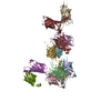
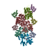
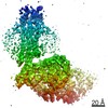
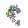
| Title | Rhodopsin-Gi complex | |||||||||||||||||||||||||||||||||||||||||||||||||||||||||||||||||||||||||||||||||
 Components Components |
| |||||||||||||||||||||||||||||||||||||||||||||||||||||||||||||||||||||||||||||||||
 Keywords Keywords | SIGNALING PROTEIN / Rhodopsin / G protein / cryo-EM / Structure | |||||||||||||||||||||||||||||||||||||||||||||||||||||||||||||||||||||||||||||||||
| Function / homology |  Function and homology information Function and homology informationOpsins / sperm head plasma membrane / rod bipolar cell differentiation / absorption of visible light / G protein-coupled opsin signaling pathway / photoreceptor inner segment membrane / podosome assembly / 11-cis retinal binding / G protein-coupled photoreceptor activity / rod photoreceptor outer segment ...Opsins / sperm head plasma membrane / rod bipolar cell differentiation / absorption of visible light / G protein-coupled opsin signaling pathway / photoreceptor inner segment membrane / podosome assembly / 11-cis retinal binding / G protein-coupled photoreceptor activity / rod photoreceptor outer segment / cellular response to light stimulus / VxPx cargo-targeting to cilium / thermotaxis / Golgi-associated vesicle membrane / detection of temperature stimulus involved in thermoception / response to light intensity / photoreceptor cell maintenance / G-protein activation / Activation of the phototransduction cascade / Glucagon-type ligand receptors / Thromboxane signalling through TP receptor / Sensory perception of sweet, bitter, and umami (glutamate) taste / G beta:gamma signalling through PI3Kgamma / G beta:gamma signalling through CDC42 / Cooperation of PDCL (PhLP1) and TRiC/CCT in G-protein beta folding / Activation of G protein gated Potassium channels / Inhibition of voltage gated Ca2+ channels via Gbeta/gamma subunits / Ca2+ pathway / G alpha (z) signalling events / High laminar flow shear stress activates signaling by PIEZO1 and PECAM1:CDH5:KDR in endothelial cells / Glucagon-like Peptide-1 (GLP1) regulates insulin secretion / Vasopressin regulates renal water homeostasis via Aquaporins / Adrenaline,noradrenaline inhibits insulin secretion / ADP signalling through P2Y purinoceptor 12 / G alpha (q) signalling events / G alpha (i) signalling events / Thrombin signalling through proteinase activated receptors (PARs) / Activation of G protein gated Potassium channels / G-protein activation / G beta:gamma signalling through PI3Kgamma / Prostacyclin signalling through prostacyclin receptor / G beta:gamma signalling through PLC beta / ADP signalling through P2Y purinoceptor 1 / Thromboxane signalling through TP receptor / Presynaptic function of Kainate receptors / G beta:gamma signalling through CDC42 / Inhibition of voltage gated Ca2+ channels via Gbeta/gamma subunits / G alpha (12/13) signalling events / Glucagon-type ligand receptors / G beta:gamma signalling through BTK / ADP signalling through P2Y purinoceptor 12 / Adrenaline,noradrenaline inhibits insulin secretion / Cooperation of PDCL (PhLP1) and TRiC/CCT in G-protein beta folding / Ca2+ pathway / Thrombin signalling through proteinase activated receptors (PARs) / G alpha (z) signalling events / Extra-nuclear estrogen signaling / ciliary membrane / photoreceptor outer segment membrane / G alpha (s) signalling events / G alpha (q) signalling events / spectrin binding / G alpha (i) signalling events / Glucagon-like Peptide-1 (GLP1) regulates insulin secretion / High laminar flow shear stress activates signaling by PIEZO1 and PECAM1:CDH5:KDR in endothelial cells / Vasopressin regulates renal water homeostasis via Aquaporins / The canonical retinoid cycle in rods (twilight vision) / alkylglycerophosphoethanolamine phosphodiesterase activity / phototransduction, visible light / phototransduction / photoreceptor outer segment / adenylate cyclase inhibitor activity / positive regulation of protein localization to cell cortex / Adenylate cyclase inhibitory pathway / T cell migration / D2 dopamine receptor binding / response to prostaglandin E / adenylate cyclase regulator activity / G protein-coupled serotonin receptor binding / adenylate cyclase-inhibiting serotonin receptor signaling pathway / sperm midpiece / cardiac muscle cell apoptotic process / photoreceptor inner segment / visual perception / cellular response to forskolin / regulation of mitotic spindle organization / Regulation of insulin secretion / positive regulation of cholesterol biosynthetic process / negative regulation of insulin secretion / electron transport chain / G protein-coupled receptor binding / G protein-coupled receptor activity / response to peptide hormone / adenylate cyclase-inhibiting G protein-coupled receptor signaling pathway / adenylate cyclase-modulating G protein-coupled receptor signaling pathway / G-protein beta/gamma-subunit complex binding / centriolar satellite / microtubule cytoskeleton organization / Activation of the phototransduction cascade / ADP signalling through P2Y purinoceptor 12 Similarity search - Function | |||||||||||||||||||||||||||||||||||||||||||||||||||||||||||||||||||||||||||||||||
| Biological species |   Homo sapiens (human) Homo sapiens (human) synthetic construct (others) | |||||||||||||||||||||||||||||||||||||||||||||||||||||||||||||||||||||||||||||||||
| Method | ELECTRON MICROSCOPY / single particle reconstruction / cryo EM / Resolution: 4.5 Å | |||||||||||||||||||||||||||||||||||||||||||||||||||||||||||||||||||||||||||||||||
 Authors Authors | Kang, Y. / Kuybeda, O. / de Waal, P.W. / Mukherjee, S. / Van Eps, N. / Dutka, P. / Zhou, X.E. / Bartesaghi, A. / Erramilli, S. / Morizumi, T. ...Kang, Y. / Kuybeda, O. / de Waal, P.W. / Mukherjee, S. / Van Eps, N. / Dutka, P. / Zhou, X.E. / Bartesaghi, A. / Erramilli, S. / Morizumi, T. / Gu, X. / Yin, Y. / Liu, P. / Jiang, Y. / Meng, X. / Zhao, G. / Melcher, K. / Earnst, O.P. / Kossiakoff, A.A. / Subramaniam, S. / Xu, H.E. | |||||||||||||||||||||||||||||||||||||||||||||||||||||||||||||||||||||||||||||||||
| Funding support |  United States, United States,  China, 4items China, 4items
| |||||||||||||||||||||||||||||||||||||||||||||||||||||||||||||||||||||||||||||||||
 Citation Citation | Journal: Nature / Year: 2018 Title: Cryo-EM structure of human rhodopsin bound to an inhibitory G protein. Authors: Yanyong Kang / Oleg Kuybeda / Parker W de Waal / Somnath Mukherjee / Ned Van Eps / Przemyslaw Dutka / X Edward Zhou / Alberto Bartesaghi / Satchal Erramilli / Takefumi Morizumi / Xin Gu / ...Authors: Yanyong Kang / Oleg Kuybeda / Parker W de Waal / Somnath Mukherjee / Ned Van Eps / Przemyslaw Dutka / X Edward Zhou / Alberto Bartesaghi / Satchal Erramilli / Takefumi Morizumi / Xin Gu / Yanting Yin / Ping Liu / Yi Jiang / Xing Meng / Gongpu Zhao / Karsten Melcher / Oliver P Ernst / Anthony A Kossiakoff / Sriram Subramaniam / H Eric Xu /   Abstract: G-protein-coupled receptors comprise the largest family of mammalian transmembrane receptors. They mediate numerous cellular pathways by coupling with downstream signalling transducers, including the ...G-protein-coupled receptors comprise the largest family of mammalian transmembrane receptors. They mediate numerous cellular pathways by coupling with downstream signalling transducers, including the hetrotrimeric G proteins G (stimulatory) and G (inhibitory) and several arrestin proteins. The structural mechanisms that define how G-protein-coupled receptors selectively couple to a specific type of G protein or arrestin remain unknown. Here, using cryo-electron microscopy, we show that the major interactions between activated rhodopsin and G are mediated by the C-terminal helix of the G α-subunit, which is wedged into the cytoplasmic cavity of the transmembrane helix bundle and directly contacts the amino terminus of helix 8 of rhodopsin. Structural comparisons of inactive, G-bound and arrestin-bound forms of rhodopsin with inactive and G-bound forms of the β-adrenergic receptor provide a foundation to understand the unique structural signatures that are associated with the recognition of G, G and arrestin by activated G-protein-coupled receptors. | |||||||||||||||||||||||||||||||||||||||||||||||||||||||||||||||||||||||||||||||||
| History |
|
- Structure visualization
| Movie |
Movie viewer |
|---|---|
| Structure viewer | Molecule: MolmilJmol/JSmol |
- Downloads & links
-Download
| PDBx/mmCIF format | 6cmo.cif.gz | 287.5 KB | Display | PDBx/mmCIF format |
|---|---|---|---|---|
| PDB format | pdb6cmo.ent.gz | 226.3 KB | Display | PDB format |
| PDBx/mmJSON format | 6cmo.json.gz | Tree view | PDBx/mmJSON format | |
| Others |  Other downloads Other downloads |
-Validation report
| Summary document | 6cmo_validation.pdf.gz | 838.7 KB | Display | wwPDB validaton report |
|---|---|---|---|---|
| Full document | 6cmo_full_validation.pdf.gz | 846.4 KB | Display | |
| Data in XML | 6cmo_validation.xml.gz | 41.9 KB | Display | |
| Data in CIF | 6cmo_validation.cif.gz | 66.9 KB | Display | |
| Arichive directory | https://data.pdbj.org/pub/pdb/validation_reports/cm/6cmoftp://data.pdbj.org/pub/pdb/validation_reports/cm/6cmo | HTTPS FTP |
-Related structure data
| Related structure data | 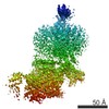 7517MC M: map data used to model this data C: citing same article ( |
|---|---|
| Similar structure data |
-Links
 PDBj
PDBj


- Assembly
| Deposited unit | 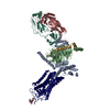
|
|---|---|
| 1 |
|
-Components
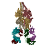
-Guanine nucleotide-binding protein ... , 3 types, 3 molecules ABG
| #2: Protein | Mass: 40445.059 Da / Num. of mol.: 1 Source method: isolated from a genetically manipulated source Source: (gene. exp.) Homo sapiens (human) / Gene: GNAI1 / Production host:   Spodoptera frugiperda (fall armyworm) / References: UniProt: P63096 Spodoptera frugiperda (fall armyworm) / References: UniProt: P63096 |
|---|---|
| #3: Protein | Mass: 37915.496 Da / Num. of mol.: 1 Source method: isolated from a genetically manipulated source Source: (gene. exp.) Spodoptera frugiperda (fall armyworm) / References: UniProt: P54311 |
| #4: Protein | Mass: 7563.750 Da / Num. of mol.: 1 Source method: isolated from a genetically manipulated source Source: (gene. exp.) Spodoptera frugiperda (fall armyworm) / References: UniProt: P63212 |
-Antibody , 2 types, 2 molecules LH
| #5: Antibody | Mass: 23258.783 Da / Num. of mol.: 1 Source method: isolated from a genetically manipulated source Source: (gene. exp.) synthetic construct (others) Production host: Bacteria Latreille et al. 1825 (Bacteria stick insect) |
|---|---|
| #6: Antibody | Mass: 25788.822 Da / Num. of mol.: 1 Source method: isolated from a genetically manipulated source Source: (gene. exp.) synthetic construct (others) Production host: Bacteria Latreille et al. 1825 (Bacteria stick insect) |
-Protein / Sugars , 2 types, 2 molecules R
| #1: Protein | Mass: 52385.406 Da / Num. of mol.: 1 Source method: isolated from a genetically manipulated source Source: (gene. exp.) Homo sapiens (human)Gene: cybC, RHO, OPN2 / Production host: Spodoptera frugiperda (fall armyworm) / References: UniProt: P0ABE7, UniProt: P08100 |
|---|---|
| #7: Polysaccharide | 2-acetamido-2-deoxy-beta-D-glucopyranose-(1-4)-2-acetamido-2-deoxy-beta-D-glucopyranose Source method: isolated from a genetically manipulated source |
-Details
| Has protein modification | Y |
|---|
-Experimental details
-Experiment
| Experiment | Method: ELECTRON MICROSCOPY |
|---|---|
| EM experiment | Aggregation state: PARTICLE / 3D reconstruction method: single particle reconstruction |
- Sample preparation
| Component | Name: Protein complex of Rhodopsin with Galphai / Type: COMPLEX / Entity ID: #1-#6 / Source: RECOMBINANT |
|---|---|
| Molecular weight | Value: 180 kDa/nm / Experimental value: YES |
| Source (natural) | Organism: Homo sapiens (human) |
| Source (recombinant) | Organism: Homo sapiens (human) |
| Buffer solution | pH: 7.2 |
| Specimen | Conc.: 9 mg/ml / Embedding applied: NO / Shadowing applied: NO / Staining applied: NO / Vitrification applied: YES |
| Vitrification | Cryogen name: ETHANE |
- Electron microscopy imaging
| Experimental equipment |  Model: Titan Krios / Image courtesy: FEI Company |
|---|---|
| Microscopy | Model: FEI TITAN KRIOS |
| Electron gun | Electron source:  FIELD EMISSION GUN / Accelerating voltage: 300 kV / Illumination mode: FLOOD BEAM FIELD EMISSION GUN / Accelerating voltage: 300 kV / Illumination mode: FLOOD BEAM |
| Electron lens | Mode: BRIGHT FIELD |
| Image recording | Electron dose: 1.92 e/Å2 / Film or detector model: GATAN K2 SUMMIT (4k x 4k) |
- Processing
| Software | Name: PHENIX / Version: 1.11.1-2575_1692: / Classification: refinement | ||||||||||||||||||||||||
|---|---|---|---|---|---|---|---|---|---|---|---|---|---|---|---|---|---|---|---|---|---|---|---|---|---|
| EM software | Name: PHENIX / Category: model refinement | ||||||||||||||||||||||||
| CTF correction | Type: NONE | ||||||||||||||||||||||||
| 3D reconstruction | Resolution: 4.5 Å / Resolution method: FSC 0.143 CUT-OFF / Num. of particles: 227386 / Symmetry type: POINT | ||||||||||||||||||||||||
| Refine LS restraints |
|
