 Movie
Movie Controller


+ Open data

- Basic information
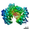
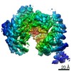
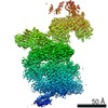
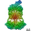
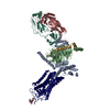
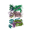
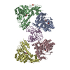
| Entry | Database: PDB / ID: 6p7x | |||||||||
|---|---|---|---|---|---|---|---|---|---|---|
| Title | Structure of the K. lactis CBF3 core - Ndc10 D1D2 complex | |||||||||
 Components Components |
| |||||||||
 Keywords Keywords | DNA BINDING PROTEIN / yeast centromere-binding complex | |||||||||
| Function / homology |  Function and homology information Function and homology informationDNA-binding transcription activator activity, RNA polymerase II-specific / ubiquitin-dependent protein catabolic process / RNA polymerase II cis-regulatory region sequence-specific DNA binding / protein ubiquitination / DNA binding / zinc ion binding / nucleus Similarity search - Function | |||||||||
| Biological species |  Kluyveromyces lactis (yeast) Kluyveromyces lactis (yeast) | |||||||||
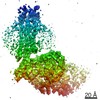
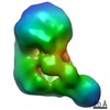
| Method | ELECTRON MICROSCOPY / single particle reconstruction / cryo EM / Resolution: 4.3 Å | |||||||||
 Authors Authors | Lee, P.D. / Wei, H. / Tan, D. / Harrison, S.C. | |||||||||
| Funding support |  United States, 2items United States, 2items
| |||||||||
 Citation Citation | Journal: J Mol Biol / Year: 2019 Title: Structure of the Centromere Binding Factor 3 Complex from Kluyveromyces lactis. Authors: Phong D Lee / Hui Wei / Dongyan Tan / Stephen C Harrison / Abstract: Kinetochores are the multiprotein complexes that link chromosomal centromeres to mitotic-spindle microtubules. Budding yeast centromeres comprise three sequential "centromere-determining elements", ...Kinetochores are the multiprotein complexes that link chromosomal centromeres to mitotic-spindle microtubules. Budding yeast centromeres comprise three sequential "centromere-determining elements", CDEI, II, and III. CDEI (8 bp) and CDEIII (∼25 bp) are conserved between Kluyveromyces lactis and Saccharomyces cerevisiae, but CDEII in the former is twice as long (160 bp) as CDEII in the latter (80 bp). The CBF3 complex recognizes CDEIII and is required for assembly of a centromeric nucleosome, which in turn recruits other kinetochore components. To understand differences in centromeric nucleosome assembly between K. lactis and S. cerevisiae, we determined the structure of a K. lactis CBF3 complex by electron cryomicroscopy at ∼4 Å resolution and compared it with published structures of S. cerevisiae CBF3. We show differences in the pose of Ndc10 and discuss potential models of the K. lactis centromeric nucleosome that account for the extended CDEII length. | |||||||||
| History |
|
- Structure visualization
| Movie |
Movie viewer |
|---|---|
| Structure viewer | Molecule: MolmilJmol/JSmol |
- Downloads & links
-Download
| PDBx/mmCIF format | 6p7x.cif.gz | 709.9 KB | Display | PDBx/mmCIF format |
|---|---|---|---|---|
| PDB format | pdb6p7x.ent.gz | 590.3 KB | Display | PDB format |
| PDBx/mmJSON format | 6p7x.json.gz | Tree view | PDBx/mmJSON format | |
| Others |  Other downloads Other downloads |
-Validation report
| Arichive directory | https://data.pdbj.org/pub/pdb/validation_reports/p7/6p7xftp://data.pdbj.org/pub/pdb/validation_reports/p7/6p7x | HTTPS FTP |
|---|
-Related structure data
| Related structure data | 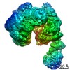 20272MC 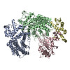 6p7vC 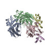 6p7wC M: map data used to model this data C: citing same article ( |
|---|---|
| Similar structure data |
-Links
 PDBj
PDBj


- Assembly
| Deposited unit | 
|
|---|---|
| 1 |
|
-Components
| #1: Protein | Mass: 47053.719 Da / Num. of mol.: 1 Source method: isolated from a genetically manipulated source Source: (gene. exp.) Kluyveromyces lactis (yeast) / Gene: KLLA0_E03807g / Production host:  |
|---|---|
| #2: Protein | Mass: 46159.945 Da / Num. of mol.: 1 Source method: isolated from a genetically manipulated source Source: (gene. exp.) Kluyveromyces lactis (yeast) / Gene: KLLA0_F13816g / Production host:  Trichoplusia ni (cabbage looper) / References: UniProt: Q6CK37 Trichoplusia ni (cabbage looper) / References: UniProt: Q6CK37 |
| #3: Protein | Mass: 21081.141 Da / Num. of mol.: 1 Source method: isolated from a genetically manipulated source Source: (gene. exp.) Kluyveromyces lactis (yeast) / Gene: SKP1 / Production host: Trichoplusia ni (cabbage looper) / References: UniProt: O94228 |
| #4: Protein | Mass: 74165.312 Da / Num. of mol.: 2 Source method: isolated from a genetically manipulated source Source: (gene. exp.) Kluyveromyces lactis (yeast) / Gene: KLLA0_D09977g / Production host: Trichoplusia ni (cabbage looper) / References: UniProt: Q6CRD4 |
-Experimental details
-Experiment
| Experiment | Method: ELECTRON MICROSCOPY |
|---|---|
| EM experiment | Aggregation state: PARTICLE / 3D reconstruction method: single particle reconstruction |
- Sample preparation
| Component | Name: K. lactis CBF3 core - Ndc10 D1D2 Complex / Type: COMPLEX / Entity ID: all / Source: RECOMBINANT |
|---|---|
| Molecular weight | Experimental value: NO |
| Source (natural) | Organism: Kluyveromyces lactis (yeast) |
| Source (recombinant) | Organism: Trichoplusia ni (cabbage looper) |
| Buffer solution | pH: 8 |
| Specimen | Embedding applied: NO / Shadowing applied: NO / Staining applied: NO / Vitrification applied: YES |
| Vitrification | Cryogen name: ETHANE |
- Electron microscopy imaging
| Experimental equipment |  Model: Titan Krios / Image courtesy: FEI Company |
|---|---|
| Microscopy | Model: FEI TITAN KRIOS |
| Electron gun | Electron source:  FIELD EMISSION GUN / Accelerating voltage: 300 kV / Illumination mode: FLOOD BEAM FIELD EMISSION GUN / Accelerating voltage: 300 kV / Illumination mode: FLOOD BEAM |
| Electron lens | Mode: BRIGHT FIELD |
| Image recording | Electron dose: 65 e/Å2 / Film or detector model: GATAN K2 SUMMIT (4k x 4k) |
- Processing
| CTF correction | Type: NONE |
|---|---|
| Symmetry | Point symmetry: C1 (asymmetric) |
| 3D reconstruction | Resolution: 4.3 Å / Resolution method: FSC 0.143 CUT-OFF / Num. of particles: 63177 / Symmetry type: POINT |
