 Movie
Movie Controller

[English] 日本語
 Yorodumi
- EMDB-4944: Cryo-EM 3D map of the N-terminal GFP tagged normal type Huntingtin -
+ Open data

- Basic information
| Entry | Database: EMDB / ID: EMD-4944 | |||||||||
|---|---|---|---|---|---|---|---|---|---|---|
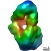
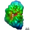
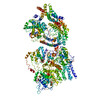
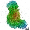
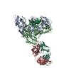
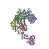
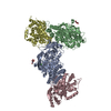
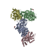
| Title | Cryo-EM 3D map of the N-terminal GFP tagged normal type Huntingtin | |||||||||
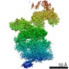
 Map data Map data | The cryo-EM map of the N-terminal GFP tagged normal Huntingtin. | |||||||||
 Sample Sample |
| |||||||||
| Biological species |  Homo sapiens (human) Homo sapiens (human) | |||||||||
| Method | single particle reconstruction / cryo EM / Resolution: 15.3 Å | |||||||||
 Authors Authors | Jung T / Tamo G / Dal Perraro M / Hebert H / Song J | |||||||||
| Funding support |  Korea, Republic Of, 2 items Korea, Republic Of, 2 items
| |||||||||
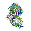
 Citation Citation | Journal: Structure / Year: 2020 Title: The Polyglutamine Expansion at the N-Terminal of Huntingtin Protein Modulates the Dynamic Configuration and Phosphorylation of the C-Terminal HEAT Domain. Authors: Taeyang Jung / Baehyun Shin / Giorgio Tamo / Hyeongju Kim / Ravi Vijayvargia / Alexander Leitner / Maria J Marcaida / Juan Astorga-Wells / Roy Jung / Ruedi Aebersold / Matteo Dal Peraro / ...Authors: Taeyang Jung / Baehyun Shin / Giorgio Tamo / Hyeongju Kim / Ravi Vijayvargia / Alexander Leitner / Maria J Marcaida / Juan Astorga-Wells / Roy Jung / Ruedi Aebersold / Matteo Dal Peraro / Hans Hebert / Ihn Sik Seong / Ji-Joon Song /    Abstract: The polyQ expansion in huntingtin protein (HTT) is the prime cause of Huntington's disease (HD). The recent cryoelectron microscopy (cryo-EM) structure of HTT-HAP40 complex provided the structural ...The polyQ expansion in huntingtin protein (HTT) is the prime cause of Huntington's disease (HD). The recent cryoelectron microscopy (cryo-EM) structure of HTT-HAP40 complex provided the structural information on its HEAT-repeat domains. Here, we present analyses of the impact of polyQ length on the structure and function of HTT via an integrative structural and biochemical approach. The cryo-EM analysis of normal (Q23) and disease (Q78) type HTTs shows that the structures of apo HTTs significantly differ from the structure of HTT in a HAP40 complex and that the polyQ expansion induces global structural changes in the relative movements among the HTT domains. In addition, we show that the polyQ expansion alters the phosphorylation pattern across HTT and that Ser2116 phosphorylation in turn affects the global structure and function of HTT. These results provide a molecular basis for the effect of the polyQ segment on HTT structure and activity, which may be important for HTT pathology. | |||||||||
| History |
|
- Structure visualization
| Movie |
 Movie viewer Movie viewer |
|---|---|
| Structure viewer | EM map: SurfViewMolmilJmol/JSmol |
| Supplemental images |

- Downloads & links
-EMDB archive
| Map data | emd_4944.map.gz | 4.9 MB | EMDB map data format | |
|---|---|---|---|---|
| Header (meta data) | emd-4944-v30.xmlemd-4944.xml | 13.7 KB 13.7 KB | Display Display | EMDB header |
| FSC (resolution estimation) | emd_4944_fsc.xml | 5.1 KB | Display | FSC data file |
| Images | 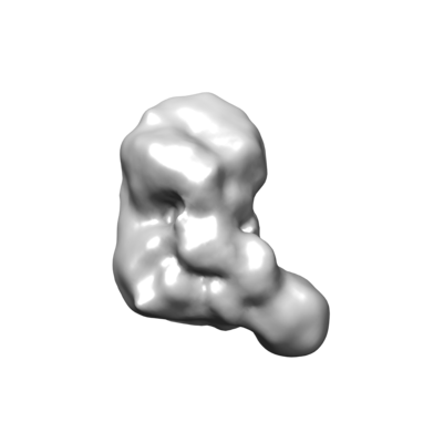 emd_4944.png emd_4944.png | 32.1 KB | ||
| Archive directory |  http://ftp.pdbj.org/pub/emdb/structures/EMD-4944ftp://ftp.pdbj.org/pub/emdb/structures/EMD-4944 http://ftp.pdbj.org/pub/emdb/structures/EMD-4944ftp://ftp.pdbj.org/pub/emdb/structures/EMD-4944 | HTTPS FTP |
-Related structure data


-Links
| EMDB pages | EMDB (EBI/PDBe) / EMDataResource |
|---|
-Map
| File | Download / File: emd_4944.map.gz / Format: CCP4 / Size: 6.6 MB / Type: IMAGE STORED AS FLOATING POINT NUMBER (4 BYTES) | ||||||||||||||||||||||||||||||||||||||||||||||||||||||||||||
|---|---|---|---|---|---|---|---|---|---|---|---|---|---|---|---|---|---|---|---|---|---|---|---|---|---|---|---|---|---|---|---|---|---|---|---|---|---|---|---|---|---|---|---|---|---|---|---|---|---|---|---|---|---|---|---|---|---|---|---|---|---|
| Annotation | The cryo-EM map of the N-terminal GFP tagged normal Huntingtin. | ||||||||||||||||||||||||||||||||||||||||||||||||||||||||||||
| Projections & slices | Image control
Images are generated by Spider. | ||||||||||||||||||||||||||||||||||||||||||||||||||||||||||||
| Voxel size | X=Y=Z: 2.20833 Å | ||||||||||||||||||||||||||||||||||||||||||||||||||||||||||||
| Density |
| ||||||||||||||||||||||||||||||||||||||||||||||||||||||||||||
| Symmetry | Space group: 1 | ||||||||||||||||||||||||||||||||||||||||||||||||||||||||||||
| Details | EMDB XML:
CCP4 map header:
| ||||||||||||||||||||||||||||||||||||||||||||||||||||||||||||
 Z (Sec.)
 Y (Row.)
 X (Col.)


-Supplemental data
- Sample components
-Entire : N-terminal GFP tagged normal type huntingtin
| Entire | Name: N-terminal GFP tagged normal type huntingtin |
|---|---|
| Components |
|
-Supramolecule #1: N-terminal GFP tagged normal type huntingtin
| Supramolecule | Name: N-terminal GFP tagged normal type huntingtin / type: organelle_or_cellular_component / ID: 1 / Parent: 0 / Macromolecule list: #1 / Details: GFP inserted at N-terminal at normal Huntingtin. |
|---|---|
| Source (natural) | Organism: Homo sapiens (human) |
| Molecular weight | Experimental: 350 KDa |
| Recombinant expression | Organism:   Spodoptera frugiperda (fall armyworm) / Recombinant cell: Sf9 cell / Recombinant plasmid: pFASTBAC1 Spodoptera frugiperda (fall armyworm) / Recombinant cell: Sf9 cell / Recombinant plasmid: pFASTBAC1 |
-Experimental details
-Structure determination
| Method | cryo EM |
|---|---|
 Processing Processing | single particle reconstruction |
| Aggregation state | particle |
-Sample preparation
| Concentration | 0.08 mg/mL | |||||||||
|---|---|---|---|---|---|---|---|---|---|---|
| Buffer | pH: 7.5 Component:
| |||||||||
| Vitrification | Cryogen name: ETHANE / Chamber humidity: 100 % / Chamber temperature: 15 K / Instrument: FEI VITROBOT MARK II / Details: 30 seconds incubation 9 seconds blotting. | |||||||||
| Details | HTT was mixed with final 0.05% of Octyl glucoside. |
- Electron microscopy
| Microscope | FEI TITAN KRIOS |
|---|---|
| Specialist optics | Phase plate: VOLTA PHASE PLATE / Energy filter - Slit width: 20 eV |
| Image recording | Film or detector model: GATAN K2 SUMMIT (4k x 4k) / Detector mode: COUNTING / Digitization - Frames/image: 2-40 / Number real images: 2226 / Average exposure time: 8.0 sec. / Average electron dose: 47.72 e/Å2 |
| Electron beam | Acceleration voltage: 300 kV / Electron source:  FIELD EMISSION GUN FIELD EMISSION GUN |
| Electron optics | Calibrated magnification: 47170 / Illumination mode: SPOT SCAN / Imaging mode: BRIGHT FIELD / Cs: 2.7 mm / Nominal defocus max: 0.8 µm / Nominal defocus min: 0.6 µm |
| Sample stage | Specimen holder model: FEI TITAN KRIOS AUTOGRID HOLDER / Cooling holder cryogen: NITROGEN |
| Experimental equipment |  Model: Titan Krios / Image courtesy: FEI Company |

