 Movie
Movie Controller

[English] 日本語
 Yorodumi
- PDB-6yej: Cryo-EM structure of the Full-length disease type human Huntingtin -
+ Open data

- Basic information
| Entry | Database: PDB / ID: 6yej | |||||||||
|---|---|---|---|---|---|---|---|---|---|---|
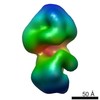
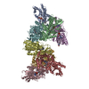
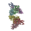
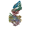
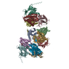
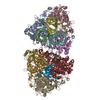
| Title | Cryo-EM structure of the Full-length disease type human Huntingtin | |||||||||
 Components Components | Huntingtin | |||||||||
 Keywords Keywords | STRUCTURAL PROTEIN / multivalent scaffold platform / PROTEIN BINDING | |||||||||
| Function / homology |  Function and homology information Function and homology informationpositive regulation of CAMKK-AMPK signaling cascade / vocal learning / regulation of CAMKK-AMPK signaling cascade / positive regulation of mitophagy / profilin binding / positive regulation of cilium assembly / retrograde vesicle-mediated transport, Golgi to endoplasmic reticulum / vesicle transport along microtubule / positive regulation of aggrephagy / positive regulation of lipophagy ...positive regulation of CAMKK-AMPK signaling cascade / vocal learning / regulation of CAMKK-AMPK signaling cascade / positive regulation of mitophagy / profilin binding / positive regulation of cilium assembly / retrograde vesicle-mediated transport, Golgi to endoplasmic reticulum / vesicle transport along microtubule / positive regulation of aggrephagy / positive regulation of lipophagy / Golgi organization / establishment of mitotic spindle orientation / dynein intermediate chain binding / synaptic vesicle transport / dynactin binding / Regulation of MECP2 expression and activity / postsynaptic cytosol / beta-tubulin binding / presynaptic cytosol / positive regulation of calcium-mediated signaling / phosphoprotein phosphatase activity / heat shock protein binding / neurogenesis / centriole / inclusion body / autophagosome / cytoplasmic vesicle membrane / negative regulation of extrinsic apoptotic signaling pathway / central nervous system development / protein destabilization / kinase binding / p53 binding / late endosome / cytoplasmic vesicle / transmembrane transporter binding / early endosome / positive regulation of apoptotic process / axon / apoptotic process / dendrite / perinuclear region of cytoplasm / Golgi apparatus / endoplasmic reticulum / protein-containing complex / nucleoplasm / identical protein binding / nucleus / cytosol / cytoplasm Similarity search - Function | |||||||||
| Biological species |  Homo sapiens (human) Homo sapiens (human) | |||||||||
| Method | ELECTRON MICROSCOPY / single particle reconstruction / cryo EM / Resolution: 18.2 Å | |||||||||
 Authors Authors | Tame, G. / Jung, T. / Dal Perraro, M. / Hebert, H. / Song, J. | |||||||||
| Funding support |  Korea, Republic Of, 2items Korea, Republic Of, 2items
| |||||||||
 Citation Citation | Journal: Structure / Year: 2020 Title: The Polyglutamine Expansion at the N-Terminal of Huntingtin Protein Modulates the Dynamic Configuration and Phosphorylation of the C-Terminal HEAT Domain. Authors: Taeyang Jung / Baehyun Shin / Giorgio Tamo / Hyeongju Kim / Ravi Vijayvargia / Alexander Leitner / Maria J Marcaida / Juan Astorga-Wells / Roy Jung / Ruedi Aebersold / Matteo Dal Peraro / ...Authors: Taeyang Jung / Baehyun Shin / Giorgio Tamo / Hyeongju Kim / Ravi Vijayvargia / Alexander Leitner / Maria J Marcaida / Juan Astorga-Wells / Roy Jung / Ruedi Aebersold / Matteo Dal Peraro / Hans Hebert / Ihn Sik Seong / Ji-Joon Song /    Abstract: The polyQ expansion in huntingtin protein (HTT) is the prime cause of Huntington's disease (HD). The recent cryoelectron microscopy (cryo-EM) structure of HTT-HAP40 complex provided the structural ...The polyQ expansion in huntingtin protein (HTT) is the prime cause of Huntington's disease (HD). The recent cryoelectron microscopy (cryo-EM) structure of HTT-HAP40 complex provided the structural information on its HEAT-repeat domains. Here, we present analyses of the impact of polyQ length on the structure and function of HTT via an integrative structural and biochemical approach. The cryo-EM analysis of normal (Q23) and disease (Q78) type HTTs shows that the structures of apo HTTs significantly differ from the structure of HTT in a HAP40 complex and that the polyQ expansion induces global structural changes in the relative movements among the HTT domains. In addition, we show that the polyQ expansion alters the phosphorylation pattern across HTT and that Ser2116 phosphorylation in turn affects the global structure and function of HTT. These results provide a molecular basis for the effect of the polyQ segment on HTT structure and activity, which may be important for HTT pathology. | |||||||||
| History |
|
- Structure visualization
| Movie |
Movie viewer |
|---|---|
| Structure viewer | Molecule: MolmilJmol/JSmol |
- Downloads & links
-Download
| PDBx/mmCIF format | 6yej.cif.gz | 380.3 KB | Display | PDBx/mmCIF format |
|---|---|---|---|---|
| PDB format | pdb6yej.ent.gz | 279.2 KB | Display | PDB format |
| PDBx/mmJSON format | 6yej.json.gz | Tree view | PDBx/mmJSON format | |
| Others |  Other downloads Other downloads |
-Validation report
| Arichive directory | https://data.pdbj.org/pub/pdb/validation_reports/ye/6yejftp://data.pdbj.org/pub/pdb/validation_reports/ye/6yej | HTTPS FTP |
|---|
-Related structure data
| Related structure data | 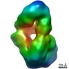 10793MC 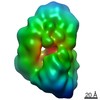 4937C 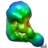 4944C 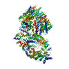 6rmhC C: citing same article ( M: map data used to model this data |
|---|---|
| Similar structure data |


-Links
 PDBj
PDBj


- Assembly
| Deposited unit | 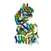
|
|---|---|
| 1 |
|
-Components
| #1: Protein | Mass: 355278.719 Da / Num. of mol.: 1 Source method: isolated from a genetically manipulated source Source: (gene. exp.) Homo sapiens (human) / Gene: HTT, HD, IT15 / Plasmid: pFASTBAC1 / Cell line (production host): Sf9 / Production host:   Spodoptera frugiperda (fall armyworm) / References: UniProt: P42858 Spodoptera frugiperda (fall armyworm) / References: UniProt: P42858 |
|---|---|
| Has protein modification | Y |
-Experimental details
-Experiment
| Experiment | Method: ELECTRON MICROSCOPY |
|---|---|
| EM experiment | Aggregation state: PARTICLE / 3D reconstruction method: single particle reconstruction |
- Sample preparation
| Component | Name: Full-length human disease-type huntingtin / Type: ORGANELLE OR CELLULAR COMPONENT Details: A disease type huntingtin with 78 polyglutamine repeat tract Entity ID: all / Source: RECOMBINANT | |||||||||||||||
|---|---|---|---|---|---|---|---|---|---|---|---|---|---|---|---|---|
| Molecular weight | Value: 0.35 MDa / Experimental value: YES | |||||||||||||||
| Source (natural) | Organism: Homo sapiens (human) | |||||||||||||||
| Source (recombinant) | Organism: Spodoptera frugiperda (fall armyworm) / Cell: Sf9 cell / Plasmid: pFASTBAC1 | |||||||||||||||
| Buffer solution | pH: 7.5 | |||||||||||||||
| Buffer component |
| |||||||||||||||
| Specimen | Conc.: 0.08 mg/ml / Embedding applied: NO / Shadowing applied: NO / Staining applied: NO / Vitrification applied: YES / Details: HTT was mixed with final 0.05% of Octyl glucoside. | |||||||||||||||
| Specimen support | Grid material: COPPER / Grid mesh size: 300 divisions/in. / Grid type: Quantifoil R2/2 | |||||||||||||||
| Vitrification | Instrument: FEI VITROBOT MARK II / Cryogen name: ETHANE / Humidity: 100 % / Chamber temperature: 288 K / Details: 30 seconds incubation 8 seconds blotting |
- Electron microscopy imaging
| Experimental equipment |  Model: Titan Krios / Image courtesy: FEI Company |
|---|---|
| Microscopy | Model: FEI TITAN KRIOS |
| Electron gun | Electron source:  FIELD EMISSION GUN / Accelerating voltage: 300 kV / Illumination mode: SPOT SCAN FIELD EMISSION GUN / Accelerating voltage: 300 kV / Illumination mode: SPOT SCAN |
| Electron lens | Mode: BRIGHT FIELD / Calibrated magnification: 47170 X / Nominal defocus max: 400 nm / Nominal defocus min: 400 nm / Cs: 2.7 mm |
| Specimen holder | Cryogen: NITROGEN / Specimen holder model: FEI TITAN KRIOS AUTOGRID HOLDER |
| Image recording | Average exposure time: 6 sec. / Electron dose: 33.6 e/Å2 / Detector mode: COUNTING / Film or detector model: GATAN K2 SUMMIT (4k x 4k) / Num. of real images: 1500 |
| EM imaging optics | Energyfilter slit width: 20 eV / Phase plate: VOLTA PHASE PLATE |
| Image scans | Movie frames/image: 24 / Used frames/image: 1-24 |
- Processing
| EM software |
| |||||||||||||||||||||||||||||||||||
|---|---|---|---|---|---|---|---|---|---|---|---|---|---|---|---|---|---|---|---|---|---|---|---|---|---|---|---|---|---|---|---|---|---|---|---|---|
| CTF correction | Type: PHASE FLIPPING AND AMPLITUDE CORRECTION | |||||||||||||||||||||||||||||||||||
| Particle selection | Num. of particles selected: 45039 / Details: manual picking | |||||||||||||||||||||||||||||||||||
| Symmetry | Point symmetry: C1 (asymmetric) | |||||||||||||||||||||||||||||||||||
| 3D reconstruction | Resolution: 18.2 Å / Resolution method: FSC 0.143 CUT-OFF / Num. of particles: 21781 / Symmetry type: POINT | |||||||||||||||||||||||||||||||||||
| Atomic model building | Protocol: RIGID BODY FIT | |||||||||||||||||||||||||||||||||||
| Atomic model building | PDB-ID: 6EZ8 Pdb chain-ID: A / Accession code: 6EZ8 / Pdb chain residue range: 91-3198 / Source name: PDB / Type: experimental model |

