[English] 日本語
Yorodumi
- PDB-3gmh: Crystal Structure of the Mad2 Dimer -

+
Open data


ID or keywords:

Loading...

-
Basic information

Entry
Database: PDB / ID: 3gmh
TitleCrystal Structure of the Mad2 Dimer
ComponentsMitotic spindle assembly checkpoint protein MAD2A
KeywordsCELL CYCLE / MITOTIC SPINDLE ASSEMBLY CHECKPOINT / Cell division / Mitosis / Nucleus
Function / homology
Function and homology information


mitotic spindle assembly checkpoint MAD1-MAD2 complex / Inhibition of the proteolytic activity of APC/C required for the onset of anaphase by mitotic spindle checkpoint components / mitotic checkpoint complex / positive regulation of mitotic cell cycle spindle assembly checkpoint / establishment of centrosome localization / Inactivation of APC/C via direct inhibition of the APC/C complex / APC/C:Cdc20 mediated degradation of mitotic proteins / nuclear pore nuclear basket / negative regulation of ubiquitin protein ligase activity / mitotic spindle assembly checkpoint signaling ...mitotic spindle assembly checkpoint MAD1-MAD2 complex / Inhibition of the proteolytic activity of APC/C required for the onset of anaphase by mitotic spindle checkpoint components / mitotic checkpoint complex / positive regulation of mitotic cell cycle spindle assembly checkpoint / establishment of centrosome localization / Inactivation of APC/C via direct inhibition of the APC/C complex / APC/C:Cdc20 mediated degradation of mitotic proteins / nuclear pore nuclear basket / negative regulation of ubiquitin protein ligase activity / mitotic spindle assembly checkpoint signaling / mitotic sister chromatid segregation / establishment of mitotic spindle orientation / negative regulation of mitotic cell cycle / Amplification of signal from unattached kinetochores via a MAD2 inhibitory signal / Mitotic Prometaphase / EML4 and NUDC in mitotic spindle formation / Resolution of Sister Chromatid Cohesion / APC-Cdc20 mediated degradation of Nek2A / RHO GTPases Activate Formins / Cdc20:Phospho-APC/C mediated degradation of Cyclin A / negative regulation of protein catabolic process / mitotic spindle / kinetochore / spindle pole / Separation of Sister Chromatids / cell division / negative regulation of apoptotic process / perinuclear region of cytoplasm / protein homodimerization activity / nucleoplasm / identical protein binding / nucleus / cytosol / cytoplasm
Similarity search - Function
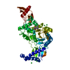
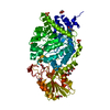
Cell Cycle, Spindle Assembly Checkpoint Protein; Chain A / HORMA domain / Mad2-like / HORMA domain / HORMA domain / HORMA domain profile. / HORMA domain superfamily / 2-Layer Sandwich / Alpha Beta
Similarity search - Domain/homology
Mitotic spindle assembly checkpoint protein MAD2A
Similarity search - Component
Biological speciesHomo sapiens (human)
MethodX-RAY DIFFRACTION / SYNCHROTRON / MOLECULAR REPLACEMENT / Resolution: 3.95 Å
AuthorsOzkan, E. / Luo, X. / Machius, M. / Yu, H. / Deisenhofer, J.
CitationJournal: Proc.Natl.Acad.Sci.USA / Year: 2015
Title: Structure of an intermediate conformer of the spindle checkpoint protein Mad2.
Authors: Hara, M. / Ozkan, E. / Sun, H. / Yu, H. / Luo, X.
History
DepositionMar 13, 2009Deposition site: RCSB / Processing site: RCSB
Revision 1.0Nov 17, 2010Provider: repository / Type: Initial release
Revision 1.1Jul 13, 2011Group: Version format compliance
Revision 1.2Sep 2, 2015Group: Database references
Revision 1.3Oct 7, 2015Group: Database references
Revision 1.4Sep 6, 2023Group: Data collection / Database references ...Data collection / Database references / Derived calculations / Refinement description
Category: chem_comp_atom / chem_comp_bond ...chem_comp_atom / chem_comp_bond / database_2 / pdbx_initial_refinement_model / struct_ref_seq_dif / struct_site
Item: _database_2.pdbx_DOI / _database_2.pdbx_database_accession ..._database_2.pdbx_DOI / _database_2.pdbx_database_accession / _struct_ref_seq_dif.details / _struct_site.pdbx_auth_asym_id / _struct_site.pdbx_auth_comp_id / _struct_site.pdbx_auth_seq_id

-
Structure visualization

Structure viewerMolecule:
MolmilJmol/JSmol

Downloads & links

-
Assembly

Deposited unit
A: Mitotic spindle assembly checkpoint protein MAD2A
B: Mitotic spindle assembly checkpoint protein MAD2A
C: Mitotic spindle assembly checkpoint protein MAD2A
D: Mitotic spindle assembly checkpoint protein MAD2A
E: Mitotic spindle assembly checkpoint protein MAD2A
F: Mitotic spindle assembly checkpoint protein MAD2A
G: Mitotic spindle assembly checkpoint protein MAD2A
H: Mitotic spindle assembly checkpoint protein MAD2A
I: Mitotic spindle assembly checkpoint protein MAD2A
J: Mitotic spindle assembly checkpoint protein MAD2A
K: Mitotic spindle assembly checkpoint protein MAD2A
L: Mitotic spindle assembly checkpoint protein MAD2A
hetero molecules


Theoretical massNumber of molelcules
Total (without water)287,04224
Polymers285,89012
Non-polymers1,15312
Water0
1
A: Mitotic spindle assembly checkpoint protein MAD2A
B: Mitotic spindle assembly checkpoint protein MAD2A
hetero molecules


Theoretical massNumber of molelcules
Total (without water)47,9365
Polymers47,6482
Non-polymers2883
Water0
TypeNameSymmetry operationNumber
identity operation1_555x,y,z1
Buried area2590 Å2
ΔGint-48 kcal/mol
Surface area19180 Å2
MethodPISA
2
C: Mitotic spindle assembly checkpoint protein MAD2A
D: Mitotic spindle assembly checkpoint protein MAD2A
hetero molecules


Theoretical massNumber of molelcules
Total (without water)47,9365
Polymers47,6482
Non-polymers2883
Water0
TypeNameSymmetry operationNumber
identity operation1_555x,y,z1
Buried area2530 Å2
ΔGint-46 kcal/mol
Surface area19790 Å2
MethodPISA
3
E: Mitotic spindle assembly checkpoint protein MAD2A
F: Mitotic spindle assembly checkpoint protein MAD2A
hetero molecules


Theoretical massNumber of molelcules
Total (without water)47,7443
Polymers47,6482
Non-polymers961
Water0
TypeNameSymmetry operationNumber
identity operation1_555x,y,z1
Buried area2240 Å2
ΔGint-22 kcal/mol
Surface area19060 Å2
MethodPISA
4
G: Mitotic spindle assembly checkpoint protein MAD2A
H: Mitotic spindle assembly checkpoint protein MAD2A
hetero molecules


Theoretical massNumber of molelcules
Total (without water)47,8404
Polymers47,6482
Non-polymers1922
Water0
TypeNameSymmetry operationNumber
identity operation1_555x,y,z1
Buried area2360 Å2
ΔGint-33 kcal/mol
Surface area19230 Å2
MethodPISA
5
I: Mitotic spindle assembly checkpoint protein MAD2A
J: Mitotic spindle assembly checkpoint protein MAD2A
hetero molecules


Theoretical massNumber of molelcules
Total (without water)47,7443
Polymers47,6482
Non-polymers961
Water0
TypeNameSymmetry operationNumber
identity operation1_555x,y,z1
Buried area2330 Å2
ΔGint-23 kcal/mol
Surface area19030 Å2
MethodPISA
6
K: Mitotic spindle assembly checkpoint protein MAD2A
L: Mitotic spindle assembly checkpoint protein MAD2A
hetero molecules


Theoretical massNumber of molelcules
Total (without water)47,8404
Polymers47,6482
Non-polymers1922
Water0
TypeNameSymmetry operationNumber
identity operation1_555x,y,z1
Buried area2310 Å2
ΔGint-35 kcal/mol
Surface area19480 Å2
MethodPISA
Unit cell
Length a, b, c (Å)124.829, 104.918, 157.746
Angle α, β, γ (deg.)90.00, 97.57, 90.00
Int Tables number4
Space group name H-MP1211
Noncrystallographic symmetry (NCS)NCS domain:
IDEns-ID
11
21
31
41
51
61
12
22
32
42
52
62
13
23

NCS domain segments:
Dom-IDComponent-IDEns-IDSelection details
111chain A and (resseq 10:202)
211chain C and (resseq 10:44 or resseq 46:204)
311chain E and (resseq 10:204)
411chain G and (resseq 10:203)
511chain I and (resseq 10:32 or resseq 44:135 or resseq 143:203)
611chain K and (resseq 10:203)
112chain B
212chain D
312chain F and (resseq 10:46 or resseq 51:198)
412chain H and (resseq 10:157 or resseq 176:203)
512chain J and not (resseq 165 and (name CB or name CG or name CD or name OE*))
612chain L and (resseq 10:157 or resseq 176:198)
113chain H and (resseq 163:172)
213chain L and (resseq 163:172)

NCS ensembles :
ID
1
2
3

-
Components

#1: Protein
Mitotic spindle assembly checkpoint protein MAD2A / MAD2-like 1 / HsMAD2


Mass: 23824.131 Da / Num. of mol.: 12
Source method: isolated from a genetically manipulated source
Source: (gene. exp.) Homo sapiens (human) / Gene: MAD2, MAD2A, MAD2L1 / Plasmid: pQE-30 / Production host: Escherichia coli (E. coli) / Strain (production host): M15[pREP4] / References: UniProt: Q13257
#2: Chemical
ChemComp-SO4 / SULFATE ION / Sulfate


Mass: 96.063 Da / Num. of mol.: 12 / Source method: obtained synthetically / Formula: SO4

-
Experimental details

-
Experiment

ExperimentMethod: X-RAY DIFFRACTION / Number of used crystals: 1

-
Sample preparation

CrystalDensity Matthews: 3.58 Å3/Da / Density % sol: 65.66 %
Crystal growTemperature: 294 K / Method: vapor diffusion, hanging drop / pH: 7.5
Details: 1.6M ammonium sulfate, 0.1M HEPES, pH 7.5, VAPOR DIFFUSION, HANGING DROP, temperature 294K

-
Data collection

DiffractionMean temperature: 120 K
Diffraction sourceSource: SYNCHROTRON / Site: APS / Beamline: 19-ID / Wavelength: 0.97929 Å
DetectorType: SBC-2 / Detector: CCD / Date: Jan 1, 2004
RadiationMonochromator: Custom / Protocol: SINGLE WAVELENGTH / Monochromatic (M) / Laue (L): M / Scattering type: x-ray
Radiation wavelengthWavelength: 0.97929 Å / Relative weight: 1
ReflectionResolution: 3.95→50 Å / Num. obs: 35718 / % possible obs: 100 % / Observed criterion σ(I): -3 / Redundancy: 7.3 % / Biso Wilson estimate: 116.12 Å2 / Rmerge(I) obs: 0.165 / Net I/σ(I): 11.6
Reflection shellResolution: 3.95→4.09 Å / Redundancy: 7.2 % / Rmerge(I) obs: 0.78 / Mean I/σ(I) obs: 2.7 / % possible all: 100

-
Processing

Software
NameVersionClassification
HKL-2000data collection
PHASERphasing
PHENIX(phenix.refine)refinement
HKL-2000data reduction
HKL-2000data scaling
RefinementMethod to determine structure: MOLECULAR REPLACEMENT
Starting model: 2V64
Resolution: 3.95→49.735 Å / SU ML: -0 / Isotropic thermal model: ISOTROPIC, WITH TLS / σ(F): 1.36 / Phase error: 24.16 / Stereochemistry target values: ML
RfactorNum. reflection% reflection
Rfree0.2505 1771 4.97 %
Rwork0.2182 --
obs0.2199 35601 99.56 %
Solvent computationShrinkage radii: 0.9 Å / VDW probe radii: 1.11 Å / Solvent model: FLAT BULK SOLVENT MODEL / Bsol: 113.136 Å2 / ksol: 0.322 e/Å3
Displacement parametersBiso max: 413.72 Å2 / Biso mean: 156.85 Å2 / Biso min: 65.1 Å2
Baniso -1Baniso -2Baniso -3
1-1.799 Å20 Å219.572 Å2
2--33.17 Å20 Å2
3----34.969 Å2
Refinement stepCycle: LAST / Resolution: 3.95→49.735 Å
ProteinNucleic acidLigandSolventTotal
Num. atoms18241 0 60 0 18301
Refine LS restraints
Refine-IDTypeDev idealNumber
X-RAY DIFFRACTIONf_bond_d0.00818620
X-RAY DIFFRACTIONf_angle_d1.02325239
X-RAY DIFFRACTIONf_dihedral_angle_d17.0966775
X-RAY DIFFRACTIONf_chiral_restr0.0683014
X-RAY DIFFRACTIONf_plane_restr0.0043118
Refine LS restraints NCS
Ens-IDDom-IDAuth asym-IDNumberRefine-IDTypeRms dev position (Å)
11A1550X-RAY DIFFRACTIONPOSITIONAL
12C1550X-RAY DIFFRACTIONPOSITIONAL0.028
13E1507X-RAY DIFFRACTIONPOSITIONAL0.039
14G1518X-RAY DIFFRACTIONPOSITIONAL0.033
15I1370X-RAY DIFFRACTIONPOSITIONAL0.04
16K1518X-RAY DIFFRACTIONPOSITIONAL0.036
21B1495X-RAY DIFFRACTIONPOSITIONAL
22D1495X-RAY DIFFRACTIONPOSITIONAL0.03
23F1414X-RAY DIFFRACTIONPOSITIONAL0.042
24H1342X-RAY DIFFRACTIONPOSITIONAL0.036
25J1490X-RAY DIFFRACTIONPOSITIONAL0.038
26L1299X-RAY DIFFRACTIONPOSITIONAL0.043
31H81X-RAY DIFFRACTIONPOSITIONAL
32L81X-RAY DIFFRACTIONPOSITIONAL0.017
LS refinement shell
Resolution (Å)Rfactor RfreeNum. reflection RfreeRfactor RworkNum. reflection RworkRefine-ID% reflection obs (%)
3.95-4.05680.32181170.27742474X-RAY DIFFRACTION95
4.0568-4.17610.25311190.24062613X-RAY DIFFRACTION100
4.1761-4.31080.25981340.22332587X-RAY DIFFRACTION100
4.3108-4.46480.2421230.20822602X-RAY DIFFRACTION100
4.4648-4.64340.2411390.19532606X-RAY DIFFRACTION100
4.6434-4.85460.21681250.19692594X-RAY DIFFRACTION100
4.8546-5.11030.21511400.18642613X-RAY DIFFRACTION100
5.1103-5.43010.2251510.19392604X-RAY DIFFRACTION100
5.4301-5.84870.23651370.2062596X-RAY DIFFRACTION100
5.8487-6.43620.2371510.21222590X-RAY DIFFRACTION100
6.4362-7.3650.23541450.2042630X-RAY DIFFRACTION100
7.365-9.26930.22171460.17952627X-RAY DIFFRACTION100
9.2693-49.73880.23821440.21432694X-RAY DIFFRACTION100
Refinement TLS params.

Refine-ID: X-RAY DIFFRACTION

IDMethodL112)L122)L132)L222)L232)L332)S11 (Å °)S12 (Å °)S13 (Å °)S21 (Å °)S22 (Å °)S23 (Å °)S31 (Å °)S32 (Å °)S33 (Å °)T112)T122)T132)T222)T232)T332)Origin x (Å)Origin y (Å)Origin z (Å)
1refined2.591.2705-0.69184.0035-1.20023.38470.12010.0356-0.4951-0.17380.0396-0.28350.358-0.2116-0.10790.84430.0678-0.39050.7762-0.24331.129693.9151-13.072755.3166
23.1794-0.4276-1.39261.9232-0.3241.72690.26190.29390.1982-0.2431-0.1746-0.3064-0.0898-0.3109-0.05120.88270.2556-0.07330.7151-0.07610.9398
33.38031.0492-0.26241.25981.84094.33890.29420.00890.7347-0.2274-0.03650.4096-0.7115-0.0686-0.33290.8794-0.08290.24650.57930.18151.354
42.1184-1.47671.89242.2504-0.28612.59250.36090.19330.0661-0.1128-0.20620.21790.01480.4245-0.05150.71910.05180.0140.7250.15190.9761
51.4272-1.3645-1.58973.5778-0.66345.14590.0755-1.33650.18750.69370.3676-0.52250.17441.9906-0.46411.0432-0.2287-0.11112.5192-0.47221.1351
63.5791-0.7555-1.99392.7617-1.03724.80.89790.53730.2903-0.57420.0894-0.22830.02590.3043-0.79031.2432-0.1290.23571.3634-0.18881.0854
72.6879-0.3220.0220.75370.30110.14790.25511.49940.0836-0.4608-0.042-0.1-0.16350.3515-0.11841.6820.1768-0.08582.12430.14261.0538
84.1350.20810.60870.09630.70634.36670.22810.11350.43540.348-0.3081-0.2379-0.5885-0.29940.08551.45510.1102-0.25060.81690.19421.1901
93.2219-1.5881-1.96352.80980.29053.7693-0.0132-0.05620.21630.29790.4828-0.0257-0.2282-0.7875-0.42031.5866-0.11190.19011.55650.21011.1546
101.7412-1.18651.69030.9917-0.52292.99950.43770.5871-0.0007-0.5833-0.07320.14440.72180.4149-0.18681.8921-0.2315-0.03111.50190.2090.9521
113.9267-0.24641.3314.27840.40250.8135-0.14592.02610.1538-0.5249-0.12960.1345-0.0051-0.00650.21831.106-0.15090.10442.2129-0.00191.0359
124.49531.0129-0.46880.13660.01085.7317-0.10610.0772-0.33390.2229-0.02730.53520.87680.66380.11931.20740.26730.2560.7679-0.08471.0947
Refinement TLS groupSelection details: chain L

+
About Yorodumi

-
News

-
Feb 9, 2022. New format data for meta-information of EMDB entries

New format data for meta-information of EMDB entries

  • Version 3 of the EMDB header file is now the official format.
  • The previous official version 1.9 will be removed from the archive.

Related info.:EMDB header

External links:wwPDB to switch to version 3 of the EMDB data model

-
Aug 12, 2020. Covid-19 info

Covid-19 info

URL: https://pdbj.org/emnavi/covid19.php

New page: Covid-19 featured information page in EM Navigator.

Related info.:Covid-19 info / Mar 5, 2020. Novel coronavirus structure data

+
Mar 5, 2020. Novel coronavirus structure data

Novel coronavirus structure data

Related info.:Yorodumi Speices / Aug 12, 2020. Covid-19 info

External links:COVID-19 featured content - PDBj / Molecule of the Month (242):Coronavirus Proteases

+
Jan 31, 2019. EMDB accession codes are about to change! (news from PDBe EMDB page)

EMDB accession codes are about to change! (news from PDBe EMDB page)

  • The allocation of 4 digits for EMDB accession codes will soon come to an end. Whilst these codes will remain in use, new EMDB accession codes will include an additional digit and will expand incrementally as the available range of codes is exhausted. The current 4-digit format prefixed with “EMD-” (i.e. EMD-XXXX) will advance to a 5-digit format (i.e. EMD-XXXXX), and so on. It is currently estimated that the 4-digit codes will be depleted around Spring 2019, at which point the 5-digit format will come into force.
  • The EM Navigator/Yorodumi systems omit the EMD- prefix.

Related info.:Q: What is EMD? / ID/Accession-code notation in Yorodumi/EM Navigator

External links:EMDB Accession Codes are Changing Soon! / Contact to PDBj

+
Jul 12, 2017. Major update of PDB

Major update of PDB

  • wwPDB released updated PDB data conforming to the new PDBx/mmCIF dictionary.
  • This is a major update changing the version number from 4 to 5, and with Remediation, in which all the entries are updated.
  • In this update, many items about electron microscopy experimental information are reorganized (e.g. em_software).
  • Now, EM Navigator and Yorodumi are based on the updated data.

External links:wwPDB Remediation / Enriched Model Files Conforming to OneDep Data Standards Now Available in the PDB FTP Archive

-
Yorodumi

Thousand views of thousand structures

  • Yorodumi is a browser for structure data from EMDB, PDB, SASBDB, etc.
  • This page is also the successor to EM Navigator detail page, and also detail information page/front-end page for Omokage search.
  • The word "yorodu" (or yorozu) is an old Japanese word meaning "ten thousand". "mi" (miru) is to see.

Related info.:EMDB / PDB / SASBDB / Comparison of 3 databanks / Yorodumi Search / Aug 31, 2016. New EM Navigator & Yorodumi / Yorodumi Papers / Jmol/JSmol / Function and homology information / Changes in new EM Navigator and Yorodumi

Read more