 Movie
Movie Controller

[English] 日本語
 Yorodumi
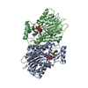
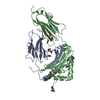
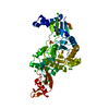
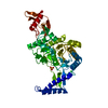
- PDB-1z6z: Crystal Structure of Human Sepiapterin Reductase in complex with NADP+ -
+ Open data

- Basic information
| Entry | Database: PDB / ID: 1z6z | ||||||
|---|---|---|---|---|---|---|---|
| Title | Crystal Structure of Human Sepiapterin Reductase in complex with NADP+ | ||||||
 Components Components | Sepiapterin reductase | ||||||
 Keywords Keywords | OXIDOREDUCTASE / short-chain dehydrogenase/reductase / sepiapterin reductase / human / Structural Genomics / Structural Genomics Consortium / SGC | ||||||
| Function / homology |  Function and homology information Function and homology informationsepiapterin reductase (L-erythro-7,8-dihydrobiopterin-forming) / sepiapterin reductase (NADP+) activity / tetrahydrobiopterin biosynthetic process / alcohol dehydrogenase (NADP+) activity / nitric oxide biosynthetic process / eNOS activation / Tetrahydrobiopterin (BH4) synthesis, recycling, salvage and regulation / NADP binding / mitochondrion / extracellular exosome ...sepiapterin reductase (L-erythro-7,8-dihydrobiopterin-forming) / sepiapterin reductase (NADP+) activity / tetrahydrobiopterin biosynthetic process / alcohol dehydrogenase (NADP+) activity / nitric oxide biosynthetic process / eNOS activation / Tetrahydrobiopterin (BH4) synthesis, recycling, salvage and regulation / NADP binding / mitochondrion / extracellular exosome / nucleoplasm / cytosol Similarity search - Function | ||||||
| Biological species |  Homo sapiens (human) Homo sapiens (human) | ||||||
| Method |  X-RAY DIFFRACTION / MOLECULAR REPLACEMENT / Resolution: 2.5 Å X-RAY DIFFRACTION / MOLECULAR REPLACEMENT / Resolution: 2.5 Å | ||||||
 Authors Authors | Ugochukwu, E. / Kavanagh, K. / Ng, S. / Arrowsmith, C. / Edwards, A. / Sundstrom, M. / von Delft, F. / Oppermann, U. / Structural Genomics Consortium (SGC) | ||||||
 Citation Citation | Journal: To be Published Title: Crystal Structure of Human Sepiapterin Reductase Authors: Ugochukwu, E. / Kavanagh, K. / Ng, S. / Arrowsmith, C. / Edwards, A. / Sundstrom, M. / von Delft, F. / Oppermann, U. | ||||||
| History |
|
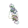
- Structure visualization
| Structure viewer | Molecule: MolmilJmol/JSmol |
|---|
- Downloads & links
-Download
| PDBx/mmCIF format | 1z6z.cif.gz | 303.3 KB | Display | PDBx/mmCIF format |
|---|---|---|---|---|
| PDB format | pdb1z6z.ent.gz | 244.7 KB | Display | PDB format |
| PDBx/mmJSON format | 1z6z.json.gz | Tree view | PDBx/mmJSON format | |
| Others |  Other downloads Other downloads |
-Validation report
| Arichive directory | https://data.pdbj.org/pub/pdb/validation_reports/z6/1z6zftp://data.pdbj.org/pub/pdb/validation_reports/z6/1z6z | HTTPS FTP |
|---|
-Related structure data
| Related structure data |  1sepS S: Starting model for refinement |
|---|---|
| Similar structure data |


-Links
 PDBj
PDBj


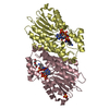
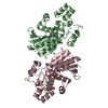
- Assembly
| Deposited unit | 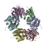
| ||||||||||||||||||||||||||||||||||||||||||||||||||||||||||||||||||||||||||||||||||||||||||||||||||||||||||||||||||||||||||||||||||||||||||||||||||||||||||||||||||||||||||||||||||||||||||||||||||||||||||||||||||||||||||||||||||||||||||||||||||||||||||||||||||||||||||||||||||||||||||||||||||||||||||
|---|---|---|---|---|---|---|---|---|---|---|---|---|---|---|---|---|---|---|---|---|---|---|---|---|---|---|---|---|---|---|---|---|---|---|---|---|---|---|---|---|---|---|---|---|---|---|---|---|---|---|---|---|---|---|---|---|---|---|---|---|---|---|---|---|---|---|---|---|---|---|---|---|---|---|---|---|---|---|---|---|---|---|---|---|---|---|---|---|---|---|---|---|---|---|---|---|---|---|---|---|---|---|---|---|---|---|---|---|---|---|---|---|---|---|---|---|---|---|---|---|---|---|---|---|---|---|---|---|---|---|---|---|---|---|---|---|---|---|---|---|---|---|---|---|---|---|---|---|---|---|---|---|---|---|---|---|---|---|---|---|---|---|---|---|---|---|---|---|---|---|---|---|---|---|---|---|---|---|---|---|---|---|---|---|---|---|---|---|---|---|---|---|---|---|---|---|---|---|---|---|---|---|---|---|---|---|---|---|---|---|---|---|---|---|---|---|---|---|---|---|---|---|---|---|---|---|---|---|---|---|---|---|---|---|---|---|---|---|---|---|---|---|---|---|---|---|---|---|---|---|---|---|---|---|---|---|---|---|---|---|---|---|---|---|---|---|---|---|---|---|---|---|---|---|---|---|---|---|---|---|---|---|---|---|---|---|---|---|---|---|---|---|---|---|---|---|---|---|---|
| 1 | 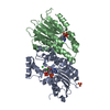
| ||||||||||||||||||||||||||||||||||||||||||||||||||||||||||||||||||||||||||||||||||||||||||||||||||||||||||||||||||||||||||||||||||||||||||||||||||||||||||||||||||||||||||||||||||||||||||||||||||||||||||||||||||||||||||||||||||||||||||||||||||||||||||||||||||||||||||||||||||||||||||||||||||||||||||
| 2 | 
| ||||||||||||||||||||||||||||||||||||||||||||||||||||||||||||||||||||||||||||||||||||||||||||||||||||||||||||||||||||||||||||||||||||||||||||||||||||||||||||||||||||||||||||||||||||||||||||||||||||||||||||||||||||||||||||||||||||||||||||||||||||||||||||||||||||||||||||||||||||||||||||||||||||||||||
| 3 | 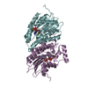
| ||||||||||||||||||||||||||||||||||||||||||||||||||||||||||||||||||||||||||||||||||||||||||||||||||||||||||||||||||||||||||||||||||||||||||||||||||||||||||||||||||||||||||||||||||||||||||||||||||||||||||||||||||||||||||||||||||||||||||||||||||||||||||||||||||||||||||||||||||||||||||||||||||||||||||
| Unit cell |
| ||||||||||||||||||||||||||||||||||||||||||||||||||||||||||||||||||||||||||||||||||||||||||||||||||||||||||||||||||||||||||||||||||||||||||||||||||||||||||||||||||||||||||||||||||||||||||||||||||||||||||||||||||||||||||||||||||||||||||||||||||||||||||||||||||||||||||||||||||||||||||||||||||||||||||
| Noncrystallographic symmetry (NCS) | NCS domain:
NCS domain segments:
|