 Movie
Movie Controller

[English] 日本語
 Yorodumi
- PDB-2je5: STRUCTURAL AND MECHANISTIC BASIS OF PENICILLIN BINDING PROTEIN IN... -
+ Open data

- Basic information
| Entry | Database: PDB / ID: 2je5 | ||||||
|---|---|---|---|---|---|---|---|
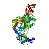
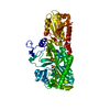
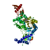
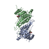
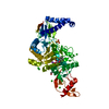
| Title | STRUCTURAL AND MECHANISTIC BASIS OF PENICILLIN BINDING PROTEIN INHIBITION BY LACTIVICINS | ||||||
 Components Components | PENICILLIN-BINDING PROTEIN 1B | ||||||
 Keywords Keywords | DRUG-BINDING PROTEIN / PEPTIDOGLYCAN SYNTHESIS MULTIFUNCTIONAL ENZYME / CELL WALL / PEPTIDOGLYCAN / GAMMA LACTAM ANTIBIOTICS | ||||||
| Function / homology |  Function and homology information Function and homology informationpeptidoglycan glycosyltransferase activity / serine-type D-Ala-D-Ala carboxypeptidase activity / penicillin binding / peptidoglycan biosynthetic process / cell wall organization / regulation of cell shape / outer membrane-bounded periplasmic space / proteolysis Similarity search - Function | ||||||
| Biological species |   STREPTOCOCCUS PNEUMONIAE (bacteria) STREPTOCOCCUS PNEUMONIAE (bacteria) | ||||||
| Method |  X-RAY DIFFRACTION / SYNCHROTRON / MOLECULAR REPLACEMENT / Resolution: 2.6 Å X-RAY DIFFRACTION / SYNCHROTRON / MOLECULAR REPLACEMENT / Resolution: 2.6 Å | ||||||
 Authors Authors | Macheboeuf, P. / Fisher, D.S. / Brown, T.J. / Zervosen, A. / Luxen, A. / Joris, B. / Dessen, A. / Schofield, C.J. | ||||||
 Citation Citation | Journal: Nat.Chem.Biol. / Year: 2007 Title: Structural and Mechanistic Basis of Penicillin-Binding Protein Inhibition by Lactivicins Authors: Macheboeuf, P. / Fisher, D.S. / Brown, T.J. / Zervosen, A. / Luxen, A. / Joris, B. / Dessen, A. / Schofield, C.J. | ||||||
| History |
|
- Structure visualization
| Structure viewer | Molecule: MolmilJmol/JSmol |
|---|
- Downloads & links
-Download
| PDBx/mmCIF format | 2je5.cif.gz | 200.2 KB | Display | PDBx/mmCIF format |
|---|---|---|---|---|
| PDB format | pdb2je5.ent.gz | 154.6 KB | Display | PDB format |
| PDBx/mmJSON format | 2je5.json.gz | Tree view | PDBx/mmJSON format | |
| Others |  Other downloads Other downloads |
-Validation report
| Arichive directory | https://data.pdbj.org/pub/pdb/validation_reports/je/2je5ftp://data.pdbj.org/pub/pdb/validation_reports/je/2je5 | HTTPS FTP |
|---|
-Related structure data
| Related structure data |  2jchC  2bg1S  2bg4 S: Starting model for refinement C: citing same article ( |
|---|---|
| Similar structure data |


-Links
 PDBj
PDBj- Assembly
| Deposited unit | 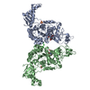
| ||||||||
|---|---|---|---|---|---|---|---|---|---|
| 1 | 
| ||||||||
| 2 | 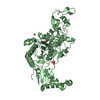
| ||||||||
| Unit cell |
| ||||||||
| Noncrystallographic symmetry (NCS) | NCS oper: (Code: given Matrix: (-0.035, -0.962, 0.27), Vector: |
-Components
| #1: Protein | Mass: 78428.328 Da / Num. of mol.: 2 / Fragment: RESIDUES 72-791 / Mutation: YES Source method: isolated from a genetically manipulated source Source: (gene. exp.) STREPTOCOCCUS PNEUMONIAE (bacteria) / Strain: R6 / Plasmid: PGEX4T1 / Production host: #2: Chemical |   Mass: 290.227 Da / Num. of mol.: 2 / Source method: obtained synthetically / Formula: C10H14N2O8 Mass: 290.227 Da / Num. of mol.: 2 / Source method: obtained synthetically / Formula: C10H14N2O8#3: Chemical |   Mass: 96.063 Da / Num. of mol.: 2 / Source method: obtained synthetically / Formula: SO4 Mass: 96.063 Da / Num. of mol.: 2 / Source method: obtained synthetically / Formula: SO4#4: Chemical | ChemComp-CL /   Mass: 35.453 Da / Num. of mol.: 4 / Source method: obtained synthetically / Formula: Cl Mass: 35.453 Da / Num. of mol.: 4 / Source method: obtained synthetically / Formula: Cl#5: Water | ChemComp-HOH / |  Mass: 18.015 Da / Num. of mol.: 79 / Source method: isolated from a natural source / Formula: H2O Mass: 18.015 Da / Num. of mol.: 79 / Source method: isolated from a natural source / Formula: H2OCompound details | ENGINEERED RESIDUE IN CHAIN A, SER 73 TO ALA ENGINEERED RESIDUE IN CHAIN A, SER 123 TO LEU ...ENGINEERED | Has protein modification | Y | |
|---|
-Experimental details
-Experiment
| Experiment | Method: X-RAY DIFFRACTION / Number of used crystals: 1 |
|---|
- Sample preparation
| Crystal | Density Matthews: 3.69 Å3/Da / Density % sol: 66.42 % / Description: NONE |
|---|---|
| Crystal grow | pH: 7 Details: 50 MH HEPES 3.2 M SODIUM CHLORIDE 0.8 M AMMONIUM SULFATE, pH 7 |
-Data collection
| Diffraction | Mean temperature: 100 K |
|---|---|
| Diffraction source | Source: SYNCHROTRON / Site: ESRF  / Beamline: ID14-2 / Wavelength: 0.933 / Beamline: ID14-2 / Wavelength: 0.933 |
| Detector | Type: ADSC CCD / Detector: CCD / Date: Feb 25, 2006 |
| Radiation | Protocol: SINGLE WAVELENGTH / Monochromatic (M) / Laue (L): M / Scattering type: x-ray |
| Radiation wavelength | Wavelength: 0.933 Å / Relative weight: 1 |
| Reflection | Resolution: 2.6→84.52 Å / Num. obs: 46529 / % possible obs: 99.2 % / Observed criterion σ(I): 5 / Redundancy: 4.7 % / Rmerge(I) obs: 0.1 / Net I/σ(I): 11.46 |
| Reflection shell | Resolution: 2.6→2.67 Å / Redundancy: 4 % / Rmerge(I) obs: 0.36 / Mean I/σ(I) obs: 4.9 / % possible all: 100 |
- Processing
| Software |
| ||||||||||||||||||||||||||||||||||||||||||||||||||||||||||||||||||||||||||||||||||||||||||||||||||||||||||||||||||||||||||||||||||||||||||||||||||||||||||||||||||||||||||||||||||||||
|---|---|---|---|---|---|---|---|---|---|---|---|---|---|---|---|---|---|---|---|---|---|---|---|---|---|---|---|---|---|---|---|---|---|---|---|---|---|---|---|---|---|---|---|---|---|---|---|---|---|---|---|---|---|---|---|---|---|---|---|---|---|---|---|---|---|---|---|---|---|---|---|---|---|---|---|---|---|---|---|---|---|---|---|---|---|---|---|---|---|---|---|---|---|---|---|---|---|---|---|---|---|---|---|---|---|---|---|---|---|---|---|---|---|---|---|---|---|---|---|---|---|---|---|---|---|---|---|---|---|---|---|---|---|---|---|---|---|---|---|---|---|---|---|---|---|---|---|---|---|---|---|---|---|---|---|---|---|---|---|---|---|---|---|---|---|---|---|---|---|---|---|---|---|---|---|---|---|---|---|---|---|---|---|
| Refinement | Method to determine structure: MOLECULAR REPLACEMENT Starting model: PDB ENTRY 2BG1 Resolution: 2.6→84.52 Å / Cor.coef. Fo:Fc: 0.943 / Cor.coef. Fo:Fc free: 0.928 / SU B: 10.87 / SU ML: 0.23 / Cross valid method: THROUGHOUT / ESU R: 0.475 / ESU R Free: 0.32 / Stereochemistry target values: MAXIMUM LIKELIHOOD / Details: HYDROGENS HAVE BEEN ADDED IN THE RIDING POSITIONS.
| ||||||||||||||||||||||||||||||||||||||||||||||||||||||||||||||||||||||||||||||||||||||||||||||||||||||||||||||||||||||||||||||||||||||||||||||||||||||||||||||||||||||||||||||||||||||
| Solvent computation | Ion probe radii: 0.8 Å / Shrinkage radii: 0.8 Å / VDW probe radii: 1.2 Å / Solvent model: MASK | ||||||||||||||||||||||||||||||||||||||||||||||||||||||||||||||||||||||||||||||||||||||||||||||||||||||||||||||||||||||||||||||||||||||||||||||||||||||||||||||||||||||||||||||||||||||
| Displacement parameters | Biso mean: 38.22 Å2
| ||||||||||||||||||||||||||||||||||||||||||||||||||||||||||||||||||||||||||||||||||||||||||||||||||||||||||||||||||||||||||||||||||||||||||||||||||||||||||||||||||||||||||||||||||||||
| Refinement step | Cycle: LAST / Resolution: 2.6→84.52 Å
| ||||||||||||||||||||||||||||||||||||||||||||||||||||||||||||||||||||||||||||||||||||||||||||||||||||||||||||||||||||||||||||||||||||||||||||||||||||||||||||||||||||||||||||||||||||||
| Refine LS restraints |
|