Movie
Movie Controller

[English] 日本語
 Yorodumi
- EMDB-6499: Structural insights into the regulatory mechanism of ATM kinase a... -
+ Open data

- Basic information
| Entry | Database: EMDB / ID: EMD-6499 | |||||||||
|---|---|---|---|---|---|---|---|---|---|---|
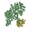
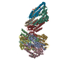
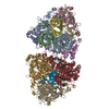
| Title | Structural insights into the regulatory mechanism of ATM kinase activation | |||||||||
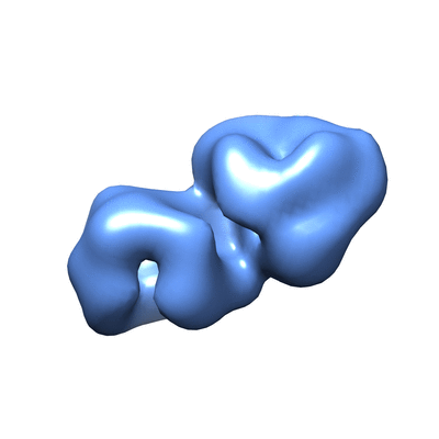
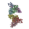
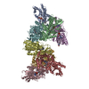
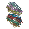
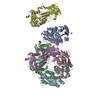
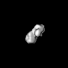
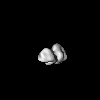
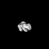
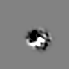
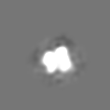
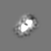
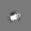
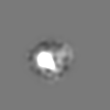
 Map data Map data | Reconstruction of monomeric ATM kinase | |||||||||
 Sample Sample |
| |||||||||
 Keywords Keywords | DNA-damage response / ataxia telangiectasia / PIKK | |||||||||
| Biological species |  Homo sapiens (human) Homo sapiens (human) | |||||||||
| Method | single particle reconstruction / negative staining / Resolution: 28.0 Å | |||||||||
 Authors Authors | Lau WCY / Li Y / Liu Z / Gao Y / Zhang Q / Huen MSY | |||||||||
 Citation Citation | Journal: Cell Cycle / Year: 2016 Title: Structure of the human dimeric ATM kinase. Authors: Wilson C Y Lau / Yinyin Li / Zhe Liu / Yuanzhu Gao / Qinfen Zhang / Michael S Y Huen /  Abstract: DNA-double strand breaks activate the serine/threonine protein kinase ataxia-telangiectasia mutated (ATM) to initiate DNA damage signal transduction. This activation process involves ...DNA-double strand breaks activate the serine/threonine protein kinase ataxia-telangiectasia mutated (ATM) to initiate DNA damage signal transduction. This activation process involves autophosphorylation and dissociation of inert ATM dimers into monomers that are catalytically active. Using single-particle electron microscopy (EM), we determined the structure of dimeric ATM in its resting state. The EM map could accommodate the crystal structure of the N-terminal truncated mammalian target of rapamycin (mTOR), a closely related enzyme of the phosphatidylinositol 3-kinase-related protein kinase (PIKK) family, allowing for the localization of the N- and the C-terminal regions of ATM. In the dimeric structure, the actives sites are buried, restricting the access of the substrates to these sites. The unanticipated domain organization of ATM provides a basis for understanding its mechanism of inhibition. | |||||||||
| History |
|
- Structure visualization
| Movie |
 Movie viewer Movie viewer |
|---|---|
| Structure viewer | EM map: SurfViewMolmilJmol/JSmol |
| Supplemental images |
- Downloads & links
-EMDB archive
| Map data | emd_6499.map.gz | 582.9 KB | EMDB map data format | |
|---|---|---|---|---|
| Header (meta data) | emd-6499-v30.xmlemd-6499.xml | 9.5 KB 9.5 KB | Display Display | EMDB header |
| Images |  400_6499.gif 400_6499.gif 80_6499.gif 80_6499.gif | 26.5 KB 2.7 KB | ||
| Archive directory |  http://ftp.pdbj.org/pub/emdb/structures/EMD-6499ftp://ftp.pdbj.org/pub/emdb/structures/EMD-6499 http://ftp.pdbj.org/pub/emdb/structures/EMD-6499ftp://ftp.pdbj.org/pub/emdb/structures/EMD-6499 | HTTPS FTP |
-Related structure data


-Links
| EMDB pages | EMDB (EBI/PDBe) / EMDataResource |
|---|
-Map
| File | Download / File: emd_6499.map.gz / Format: CCP4 / Size: 3.7 MB / Type: IMAGE STORED AS FLOATING POINT NUMBER (4 BYTES) | ||||||||||||||||||||||||||||||||||||||||||||||||||||||||||||
|---|---|---|---|---|---|---|---|---|---|---|---|---|---|---|---|---|---|---|---|---|---|---|---|---|---|---|---|---|---|---|---|---|---|---|---|---|---|---|---|---|---|---|---|---|---|---|---|---|---|---|---|---|---|---|---|---|---|---|---|---|---|
| Annotation | Reconstruction of monomeric ATM kinase | ||||||||||||||||||||||||||||||||||||||||||||||||||||||||||||
| Projections & slices | Image control
Images are generated by Spider. | ||||||||||||||||||||||||||||||||||||||||||||||||||||||||||||
| Voxel size | X=Y=Z: 4.61 Å | ||||||||||||||||||||||||||||||||||||||||||||||||||||||||||||
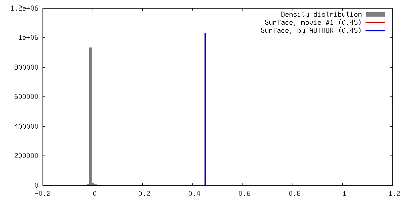
| Density |
| ||||||||||||||||||||||||||||||||||||||||||||||||||||||||||||
| Symmetry | Space group: 1 | ||||||||||||||||||||||||||||||||||||||||||||||||||||||||||||
| Details | EMDB XML:
CCP4 map header:
| ||||||||||||||||||||||||||||||||||||||||||||||||||||||||||||
 Z (Sec.)
 Y (Row.)
 X (Col.)


-Supplemental data
- Sample components
-Entire : Monomeric ATM kinase
| Entire | Name: Monomeric ATM kinase |
|---|---|
| Components |
|
-Supramolecule #1000: Monomeric ATM kinase
| Supramolecule | Name: Monomeric ATM kinase / type: sample / ID: 1000 / Details: The sample was mono disperse / Oligomeric state: Monomer / Number unique components: 1 |
|---|---|
| Molecular weight | Theoretical: 350 KDa |
-Macromolecule #1: Monomeric ATM kinase
| Macromolecule | Name: Monomeric ATM kinase / type: protein_or_peptide / ID: 1 / Number of copies: 1 / Oligomeric state: Monomer / Recombinant expression: Yes |
|---|---|
| Source (natural) | Organism: Homo sapiens (human) / synonym: Human |
| Molecular weight | Theoretical: 350 KDa |
| Recombinant expression | Organism: Homo sapiens (human) / Recombinant cell: 293T / Recombinant plasmid: pcDNA-FLAG-His-ATM wt |
-Experimental details
-Structure determination
| Method | negative staining |
|---|---|
 Processing Processing | single particle reconstruction |
| Aggregation state | particle |
-Sample preparation
| Concentration | 0.02 mg/mL |
|---|---|
| Buffer | pH: 8 / Details: 25 mM Tris, 100 mM NaCl, 1 mM TCEP, 10% glycerol |
| Staining | Type: NEGATIVE / Details: 1% w/v uranyl acetate |
| Grid | Details: Continuous carbon coated copper grids |
| Vitrification | Cryogen name: NONE / Instrument: OTHER |
- Electron microscopy
| Microscope | JEOL 2010 |
|---|---|
| Temperature | Average: 296 K |
| Date | Jul 1, 2015 |
| Image recording | Category: CCD / Film or detector model: GATAN ULTRASCAN 4000 (4k x 4k) / Number real images: 251 / Average electron dose: 25 e/Å2 |
| Electron beam | Acceleration voltage: 200 kV / Electron source: LAB6 |
| Electron optics | Illumination mode: FLOOD BEAM / Imaging mode: BRIGHT FIELD / Cs: 2.0 mm / Nominal defocus max: 4.0 µm / Nominal defocus min: 1.0 µm / Nominal magnification: 50000 |
| Sample stage | Specimen holder model: GATAN LIQUID NITROGEN |
-Image processing
| CTF correction | Details: class average |
|---|---|
| Final reconstruction | Algorithm: OTHER / Resolution.type: BY AUTHOR / Resolution: 28.0 Å / Resolution method: OTHER / Software - Name: PRIME, EMAN2 / Number images used: 6546 |
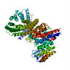
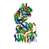
-Atomic model buiding 1
| Initial model | PDB ID: Chain - Chain ID: A |
|---|---|
| Software | Name:  Chimera Chimera |
| Details | Residues 2021-2118 are removed |
| Refinement | Space: REAL / Protocol: RIGID BODY FIT / Target criteria: cross correlation coefficient |