 Movie
Movie Controller


+ Open data

- Basic information
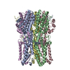
| Entry | Database: PDB / ID: 5zfp | |||||||||
|---|---|---|---|---|---|---|---|---|---|---|
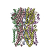
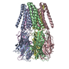
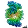
| Title | Structure of the ExbB/ExbD hexameric complex | |||||||||
 Components Components | Biopolymer transport protein ExbB | |||||||||
 Keywords Keywords | TRANSPORT PROTEIN / TON SYSTEM / ION CHANNEL / ENERGIZER / TRANSPORTER / PROTON MOTIVE FORCE | |||||||||
| Function / homology |  Function and homology information Function and homology informationferrichrome import into cell / energy transducer activity / Metal ion assimilation from the host / bacteriocin transport / cobalamin transport / intracellular monoatomic cation homeostasis / Iron assimilation using enterobactin / protein import / plasma membrane protein complex / transmembrane transporter complex ...ferrichrome import into cell / energy transducer activity / Metal ion assimilation from the host / bacteriocin transport / cobalamin transport / intracellular monoatomic cation homeostasis / Iron assimilation using enterobactin / protein import / plasma membrane protein complex / transmembrane transporter complex / Heme assimilation / transmembrane transporter activity / cell outer membrane / intracellular iron ion homeostasis / protein stabilization / membrane / identical protein binding / plasma membrane Similarity search - Function | |||||||||
| Biological species |  | |||||||||
| Method |  X-RAY DIFFRACTION / SYNCHROTRON / MOLECULAR REPLACEMENT / Resolution: 2.84 Å X-RAY DIFFRACTION / SYNCHROTRON / MOLECULAR REPLACEMENT / Resolution: 2.84 Å | |||||||||
 Authors Authors | Maki-Yonekura, S. / Matsuoka, R. / Yonekura, K. | |||||||||
| Funding support |  Japan, 2items Japan, 2items
| |||||||||
 Citation Citation | Journal: Elife / Year: 2018 Title: Hexameric and pentameric complexes of the ExbBD energizer in the Ton system. Authors: Saori Maki-Yonekura / Rei Matsuoka / Yoshiki Yamashita / Hirofumi Shimizu / Maiko Tanaka / Fumie Iwabuki / Koji Yonekura / Abstract: Gram-negative bacteria import essential nutrients such as iron and vitamin B through outer membrane receptors. This process utilizes proton motive force harvested by the Ton system made up of three ...Gram-negative bacteria import essential nutrients such as iron and vitamin B through outer membrane receptors. This process utilizes proton motive force harvested by the Ton system made up of three inner membrane proteins, ExbB, ExbD and TonB. ExbB and ExbD form the proton channel that energizes uptake through TonB. Recently, crystal structures suggest that the ExbB pentamer is the scaffold. Here, we present structures of hexameric complexes of ExbB and ExbD revealed by X-ray crystallography and single particle cryo-EM. Image analysis shows that hexameric and pentameric complexes coexist, with the proportion of hexamer increasing with pH. Channel current measurement and 2D crystallography support the existence and transition of the two oligomeric states in membranes. The hexameric complex consists of six ExbB subunits and three ExbD transmembrane helices enclosed within the central channel. We propose models for activation/inactivation associated with hexamer and pentamer formation and utilization of proton motive force. | |||||||||
| History |
|
- Structure visualization
| Structure viewer | Molecule: MolmilJmol/JSmol |
|---|
- Downloads & links
-Download
| PDBx/mmCIF format | 5zfp.cif.gz | 498.1 KB | Display | PDBx/mmCIF format |
|---|---|---|---|---|
| PDB format | pdb5zfp.ent.gz | 414 KB | Display | PDB format |
| PDBx/mmJSON format | 5zfp.json.gz | Tree view | PDBx/mmJSON format | |
| Others |  Other downloads Other downloads |
-Validation report
| Arichive directory | https://data.pdbj.org/pub/pdb/validation_reports/zf/5zfpftp://data.pdbj.org/pub/pdb/validation_reports/zf/5zfp | HTTPS FTP |
|---|
-Related structure data
| Related structure data |  6927C  6928C  5zfuC 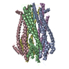 5zfvC  5sv0S S: Starting model for refinement C: citing same article ( |
|---|---|
| Similar structure data |


-Links
 PDBj
PDBj- Assembly
| Deposited unit | 
| ||||||||
|---|---|---|---|---|---|---|---|---|---|
| 1 | 
| ||||||||
| 2 | 
| ||||||||
| Unit cell |
|
-Components
| #1: Protein | Mass: 26312.322 Da / Num. of mol.: 12 Source method: isolated from a genetically manipulated source Source: (gene. exp.) Strain: K12 / Gene: exbB, b3006, JW2974 / Production host: #2: Water | ChemComp-HOH / |  Mass: 18.015 Da / Num. of mol.: 769 / Source method: isolated from a natural source / Formula: H2O Mass: 18.015 Da / Num. of mol.: 769 / Source method: isolated from a natural source / Formula: H2O |
|---|
-Experimental details
-Experiment
| Experiment | Method: X-RAY DIFFRACTION / Number of used crystals: 1 |
|---|
- Sample preparation
| Crystal | Density Matthews: 3.1 Å3/Da / Density % sol: 60.37 % |
|---|---|
| Crystal grow | Temperature: 293 K / Method: vapor diffusion, hanging drop / pH: 9 Details: 0.1 M glycine, pH 9.0, 0.15 M CaCl2, 40 % PEG 350 MME, 0.05-0.2 M L-arginine |
-Data collection
| Diffraction | Mean temperature: 90 K |
|---|---|
| Diffraction source | Source: SYNCHROTRON / Site: SPring-8 / Beamline: BL41XU / Wavelength: 1 Å |
| Detector | Type: DECTRIS PILATUS3 6M / Detector: PIXEL / Date: Oct 20, 2016 |
| Radiation | Protocol: SINGLE WAVELENGTH / Monochromatic (M) / Laue (L): M / Scattering type: x-ray |
| Radiation wavelength | Wavelength: 1 Å / Relative weight: 1 |
| Reflection | Resolution: 2.84→50.18 Å / Num. obs: 90711 / % possible obs: 99.3 % / Redundancy: 5 % / Biso Wilson estimate: 53.73 Å2 / CC1/2: 0.992 / Rmerge(I) obs: 0.11 / Rrim(I) all: 0.155 / Net I/σ(I): 6.67 |
| Reflection shell | Resolution: 2.84→2.94 Å / Rmerge(I) obs: 0.5 / CC1/2: 0.751 / Rrim(I) all: 0.706 |
- Processing
| Software |
| ||||||||||||||||||||||||||||||||||||||||||||||||||||||||||||||||||||||||||||||||||||||||||||||||||||||||||||||||||
|---|---|---|---|---|---|---|---|---|---|---|---|---|---|---|---|---|---|---|---|---|---|---|---|---|---|---|---|---|---|---|---|---|---|---|---|---|---|---|---|---|---|---|---|---|---|---|---|---|---|---|---|---|---|---|---|---|---|---|---|---|---|---|---|---|---|---|---|---|---|---|---|---|---|---|---|---|---|---|---|---|---|---|---|---|---|---|---|---|---|---|---|---|---|---|---|---|---|---|---|---|---|---|---|---|---|---|---|---|---|---|---|---|---|---|---|
| Refinement | Method to determine structure: MOLECULAR REPLACEMENT Starting model: 5SV0 Resolution: 2.84→50.18 Å / Cor.coef. Fo:Fc: 0.888 / Cor.coef. Fo:Fc free: 0.814 / Rfactor Rfree error: 0 / SU R Cruickshank DPI: 1.112 / Cross valid method: THROUGHOUT / σ(F): 0 / SU R Blow DPI: 1.049 / SU Rfree Blow DPI: 0.382 / SU Rfree Cruickshank DPI: 0.391
| ||||||||||||||||||||||||||||||||||||||||||||||||||||||||||||||||||||||||||||||||||||||||||||||||||||||||||||||||||
| Displacement parameters | Biso mean: 43.59 Å2
| ||||||||||||||||||||||||||||||||||||||||||||||||||||||||||||||||||||||||||||||||||||||||||||||||||||||||||||||||||
| Refine analyze | Luzzati coordinate error obs: 0.46 Å | ||||||||||||||||||||||||||||||||||||||||||||||||||||||||||||||||||||||||||||||||||||||||||||||||||||||||||||||||||
| Refinement step | Cycle: 1 / Resolution: 2.84→50.18 Å
| ||||||||||||||||||||||||||||||||||||||||||||||||||||||||||||||||||||||||||||||||||||||||||||||||||||||||||||||||||
| Refine LS restraints |
| ||||||||||||||||||||||||||||||||||||||||||||||||||||||||||||||||||||||||||||||||||||||||||||||||||||||||||||||||||
| LS refinement shell | Resolution: 2.84→2.91 Å / Rfactor Rfree error: 0 / Total num. of bins used: 20
|
