Movie
Movie Controller

[English] 日本語
 Yorodumi
- PDB-3jbz: Crystal structure of mTOR docked into EM map of dimeric ATM kinase -
+ Open data

- Basic information
| Entry | Database: PDB / ID: 3jbz | ||||||
|---|---|---|---|---|---|---|---|
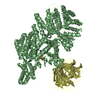
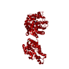
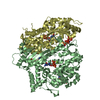
| Title | Crystal structure of mTOR docked into EM map of dimeric ATM kinase | ||||||
 Components Components | Serine/threonine-protein kinase mTOR | ||||||
 Keywords Keywords | TRANSFERASE / mTOR / PIKK | ||||||
| Function / homology |  Function and homology information Function and homology informationpositive regulation of SCF-dependent proteasomal ubiquitin-dependent catabolic process / RNA polymerase III type 2 promoter sequence-specific DNA binding / RNA polymerase III type 1 promoter sequence-specific DNA binding / positive regulation of cytoplasmic translational initiation / regulation of locomotor rhythm / T-helper 1 cell lineage commitment / positive regulation of pentose-phosphate shunt / positive regulation of wound healing, spreading of epidermal cells / regulation of membrane permeability / TORC2 complex ...positive regulation of SCF-dependent proteasomal ubiquitin-dependent catabolic process / RNA polymerase III type 2 promoter sequence-specific DNA binding / RNA polymerase III type 1 promoter sequence-specific DNA binding / positive regulation of cytoplasmic translational initiation / regulation of locomotor rhythm / T-helper 1 cell lineage commitment / positive regulation of pentose-phosphate shunt / positive regulation of wound healing, spreading of epidermal cells / regulation of membrane permeability / TORC2 complex / cellular response to leucine starvation / TFIIIC-class transcription factor complex binding / heart valve morphogenesis / voluntary musculoskeletal movement / negative regulation of lysosome organization / TORC1 complex / calcineurin-NFAT signaling cascade / positive regulation of transcription of nucleolar large rRNA by RNA polymerase I / RNA polymerase III type 3 promoter sequence-specific DNA binding / positive regulation of keratinocyte migration / regulation of osteoclast differentiation / MTOR signalling / energy reserve metabolic process / regulation of lysosome organization / cellular response to L-leucine / Energy dependent regulation of mTOR by LKB1-AMPK / cellular response to nutrient / regulation of autophagosome assembly / Amino acids regulate mTORC1 / cellular response to methionine / negative regulation of cell size / TORC2 signaling / cellular response to osmotic stress / cell projection organization / anoikis / inositol hexakisphosphate binding / cardiac muscle cell development / negative regulation of calcineurin-NFAT signaling cascade / positive regulation of ubiquitin-dependent protein catabolic process / regulation of myelination / negative regulation of protein localization to nucleus / positive regulation of transcription by RNA polymerase III / positive regulation of ruffle assembly / regulation of cell size / positive regulation of myotube differentiation / negative regulation of macroautophagy / Macroautophagy / Constitutive Signaling by AKT1 E17K in Cancer / positive regulation of actin filament polymerization / germ cell development / oligodendrocyte differentiation / TORC1 signaling / positive regulation of oligodendrocyte differentiation / behavioral response to pain / response to amino acid / positive regulation of translational initiation / TOR signaling / mTORC1-mediated signalling / CD28 dependent PI3K/Akt signaling / HSF1-dependent transactivation / regulation of macroautophagy / 'de novo' pyrimidine nucleobase biosynthetic process / positive regulation of epithelial to mesenchymal transition / positive regulation of lipid biosynthetic process / vascular endothelial cell response to laminar fluid shear stress / heart morphogenesis / regulation of cellular response to heat / positive regulation of lamellipodium assembly / neuronal action potential / T cell costimulation / phagocytic vesicle / positive regulation of stress fiber assembly / cardiac muscle contraction / negative regulation of insulin receptor signaling pathway / cytoskeleton organization / endomembrane system / cellular response to nutrient levels / positive regulation of glycolytic process / cellular response to amino acid starvation / cellular response to starvation / Regulation of PTEN gene transcription / regulation of signal transduction by p53 class mediator / negative regulation of autophagy / VEGFR2 mediated vascular permeability / post-embryonic development / TP53 Regulates Metabolic Genes / regulation of actin cytoskeleton organization / positive regulation of translation / non-specific protein-tyrosine kinase / macroautophagy / cellular response to amino acid stimulus / non-membrane spanning protein tyrosine kinase activity / phosphoprotein binding / regulation of cell growth / regulation of circadian rhythm / response to nutrient levels / PML body / protein destabilization / multicellular organism growth / response to virus Similarity search - Function | ||||||
| Biological species |  Homo sapiens (human) Homo sapiens (human) | ||||||
| Method | ELECTRON MICROSCOPY / single particle reconstruction / negative staining / Resolution: 28 Å | ||||||
 Authors Authors | Lau, W.C.Y. | ||||||
 Citation Citation | Journal: Cell Cycle / Year: 2016 Title: Structure of the human dimeric ATM kinase. Authors: Wilson C Y Lau / Yinyin Li / Zhe Liu / Yuanzhu Gao / Qinfen Zhang / Michael S Y Huen /  Abstract: DNA-double strand breaks activate the serine/threonine protein kinase ataxia-telangiectasia mutated (ATM) to initiate DNA damage signal transduction. This activation process involves ...DNA-double strand breaks activate the serine/threonine protein kinase ataxia-telangiectasia mutated (ATM) to initiate DNA damage signal transduction. This activation process involves autophosphorylation and dissociation of inert ATM dimers into monomers that are catalytically active. Using single-particle electron microscopy (EM), we determined the structure of dimeric ATM in its resting state. The EM map could accommodate the crystal structure of the N-terminal truncated mammalian target of rapamycin (mTOR), a closely related enzyme of the phosphatidylinositol 3-kinase-related protein kinase (PIKK) family, allowing for the localization of the N- and the C-terminal regions of ATM. In the dimeric structure, the actives sites are buried, restricting the access of the substrates to these sites. The unanticipated domain organization of ATM provides a basis for understanding its mechanism of inhibition. | ||||||
| History |
|
- Structure visualization
| Movie |
Movie viewer |
|---|---|
| Structure viewer | Molecule: MolmilJmol/JSmol |
 UCSF Chimera
- Downloads & links
-Download
| PDBx/mmCIF format | 3jbz.cif.gz | 212.6 KB | Display | PDBx/mmCIF format |
|---|---|---|---|---|
| PDB format | pdb3jbz.ent.gz | 160.8 KB | Display | PDB format |
| PDBx/mmJSON format | 3jbz.json.gz | Tree view | PDBx/mmJSON format | |
| Others |  Other downloads Other downloads |
-Validation report
| Arichive directory | https://data.pdbj.org/pub/pdb/validation_reports/jb/3jbzftp://data.pdbj.org/pub/pdb/validation_reports/jb/3jbz | HTTPS FTP |
|---|
-Related structure data
| Related structure data |  6501MC 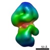 6499C M: map data used to model this data C: citing same article ( |
|---|---|
| Similar structure data |


-Links
 PDBj
PDBj


- Assembly
| Deposited unit | 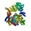
|
|---|---|
| 1 |
|
-Components
| #1: Protein | Mass: 133753.281 Da / Num. of mol.: 1 Fragment: C-terminal domain (UNP RESIDUES 1385-2020, 2119-2549) Source method: isolated from a genetically manipulated source Source: (gene. exp.) Homo sapiens (human) / Gene: FRAP, FRAP1, FRAP2, MTOR, RAFT1, RAPT1 / Cell line (production host): HEK293F / Production host: Homo sapiens (human)References: UniProt: P42345, non-specific serine/threonine protein kinase | ||
|---|---|---|---|
| #2: Chemical | ChemComp-ADP /   Mass: 427.201 Da / Num. of mol.: 1 / Source method: obtained synthetically / Formula: C10H15N5O10P2 / Comment: ADP, energy-carrying molecule*YM Mass: 427.201 Da / Num. of mol.: 1 / Source method: obtained synthetically / Formula: C10H15N5O10P2 / Comment: ADP, energy-carrying molecule*YM | ||
| #3: Chemical |   Mass: 24.305 Da / Num. of mol.: 2 / Source method: obtained synthetically / Formula: Mg Mass: 24.305 Da / Num. of mol.: 2 / Source method: obtained synthetically / Formula: Mg#4: Chemical | ChemComp-MGF / |   Mass: 81.300 Da / Num. of mol.: 1 / Source method: obtained synthetically / Formula: F3Mg Mass: 81.300 Da / Num. of mol.: 1 / Source method: obtained synthetically / Formula: F3Mg |
-Experimental details
-Experiment
| Experiment | Method: ELECTRON MICROSCOPY |
|---|---|
| EM experiment | Aggregation state: PARTICLE / 3D reconstruction method: single particle reconstruction |
- Sample preparation
| Component | Name: dimeric ATM kinase / Type: COMPLEX |
|---|---|
| Molecular weight | Value: 0.7 MDa / Experimental value: NO |
| Buffer solution | Name: 25 mM Tris pH 8.0, 100 mM NaCl, 1 mM TCEP, 10% glycerol pH: 8 / Details: 25 mM Tris, 100 mM NaCl, 1 mM TCEP, 10% glycerol |
| Specimen | Conc.: 0.02 mg/ml / Embedding applied: NO / Shadowing applied: NO / Staining applied: YES / Vitrification applied: NO |
| EM staining | Type: NEGATIVE / Details: 2% uranyl acetate / Material: uranyl acetate |
| Specimen support | Details: continuous carbon grid |
- Electron microscopy imaging
| Microscopy | Model: JEOL 2010 / Date: Sep 1, 2015 |
|---|---|
| Electron gun | Electron source: LAB6 / Accelerating voltage: 200 kV / Illumination mode: FLOOD BEAM |
| Electron lens | Mode: BRIGHT FIELD / Nominal magnification: 50000 X / Nominal defocus max: 4000 nm / Nominal defocus min: 1000 nm / Cs: 2 mm |
| Specimen holder | Temperature: 293 K |
| Image recording | Electron dose: 25 e/Å2 / Film or detector model: GATAN ULTRASCAN 4000 (4k x 4k) / Num. of real images: 251 |
| Image scans | Num. digital images: 251 |
- Processing
| EM software |
| ||||||||||||
|---|---|---|---|---|---|---|---|---|---|---|---|---|---|
| Symmetry | Point symmetry: C2 (2 fold cyclic) | ||||||||||||
| 3D reconstruction | Resolution: 28 Å / Resolution method: FSC 0.143 CUT-OFF / Symmetry type: POINT | ||||||||||||
| Atomic model building | PDB-ID: 4JSV Accession code: 4JSV / Source name: PDB / Type: experimental model | ||||||||||||
| Refinement step | Cycle: LAST
|