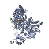
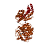
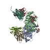
Biological unit is a homodimer. There is 1 biological unit in the asymmetric unit (chains A & B)
-
Components
#1: Protein
ProbabletRNAsulfurtransferase / Sulfur carrier protein ThiS sulfurtransferase / Thiamine biosynthesis protein ThiI / tRNA 4- ...Sulfur carrier protein ThiS sulfurtransferase / Thiamine biosynthesis protein ThiI / tRNA 4-thiouridine synthase
Mass: 44193.094 Da / Num. of mol.: 2 / Mutation: E2K Source method: isolated from a genetically manipulated source Source: (gene. exp.) Thermotoga maritima (bacteria) / Strain: ATCC 43589 / Gene: AAD36761, thiI, TM_1694 / Plasmid: pET-28a / Production host: Escherichia coli (E. coli) / Strain (production host): BL21(DE3) References: UniProt: Q9X220, tRNA uracil 4-sulfurtransferase
#2: RNA chain
RNA (39-MER)
Mass: 12559.541 Da / Num. of mol.: 2 / Source method: obtained synthetically
Mass: 24.305 Da / Num. of mol.: 2 / Source method: obtained synthetically / Formula: Mg
-
Experimental details
-
Experiment
Experiment
Method: X-RAY DIFFRACTION / Number of used crystals: 1
-
Sample preparation
Crystal
Density Matthews: 3.43 Å3/Da / Density % sol: 64.1 %
Crystal grow
Temperature: 292 K / Method: vapor diffusion, sitting drop / pH: 4.6 Details: 1 micro-liter of ThiI-RNA complex solution at 10 mg/ml in a buffer consisting of 150 mM ammonium sulfate, 20mM Tris/HCl pH 7.5 was mixed with 3 micro-liter of freshly prepared reservoir ...Details: 1 micro-liter of ThiI-RNA complex solution at 10 mg/ml in a buffer consisting of 150 mM ammonium sulfate, 20mM Tris/HCl pH 7.5 was mixed with 3 micro-liter of freshly prepared reservoir solution (2.0 M sodium formate, 100 mM sodium citrate, 2 mM DTT), VAPOR DIFFUSION, SITTING DROP, temperature 292K
In the structure databanks used in Yorodumi, some data are registered as the other names, "COVID-19 virus" and "2019-nCoV". Here are the details of the virus and the list of structure data.
Jan 31, 2019. EMDB accession codes are about to change! (news from PDBe EMDB page)
EMDB accession codes are about to change! (news from PDBe EMDB page)
The allocation of 4 digits for EMDB accession codes will soon come to an end. Whilst these codes will remain in use, new EMDB accession codes will include an additional digit and will expand incrementally as the available range of codes is exhausted. The current 4-digit format prefixed with “EMD-” (i.e. EMD-XXXX) will advance to a 5-digit format (i.e. EMD-XXXXX), and so on. It is currently estimated that the 4-digit codes will be depleted around Spring 2019, at which point the 5-digit format will come into force.
The EM Navigator/Yorodumi systems omit the EMD- prefix.
Related info.:Q: What is EMD? / ID/Accession-code notation in Yorodumi/EM Navigator
Yorodumi is a browser for structure data from EMDB, PDB, SASBDB, etc.
This page is also the successor to EM Navigator detail page, and also detail information page/front-end page for Omokage search.
The word "yorodu" (or yorozu) is an old Japanese word meaning "ten thousand". "mi" (miru) is to see.
Related info.:EMDB / PDB / SASBDB / Comparison of 3 databanks / Yorodumi Search / Aug 31, 2016. New EM Navigator & Yorodumi / Yorodumi Papers / Jmol/JSmol / Function and homology information / Changes in new EM Navigator and Yorodumi
 Movie
Movie Controller

 Yorodumi
 Open data
 Basic information
 Components
 Keywords
 Function and homology information
 Thermotoga maritima (bacteria)
 X-RAY DIFFRACTION /
  Authors
 Citation
 Structure visualization
 Downloads & links
 Other downloads
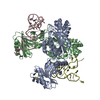


 PDBj
PDBj


 Assembly

 Mass: 507.181 Da / Num. of mol.: 2 / Source method: obtained synthetically / Formula: C10H16N5O13P3 / Comment: ATP, energy-carrying molecule*YM
 Mass: 24.305 Da / Num. of mol.: 2 / Source method: obtained synthetically / Formula: Mg
 Sample preparation
 / Beamline: X11 / Wavelength: 0.814 Å
 Processing