 Movie
Movie Controller


+ Open data

- Basic information
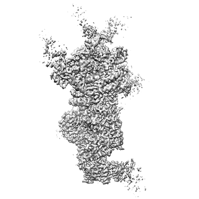
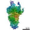
| Entry | Database: EMDB / ID: EMD-0361 | |||||||||||||||
|---|---|---|---|---|---|---|---|---|---|---|---|---|---|---|---|---|
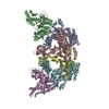
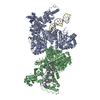
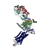
| Title | Saccharomyces cerevisiae spliceosomal E complex (ACT1) | |||||||||||||||
 Map data Map data | Spliceosomal E complex (ACT1) | |||||||||||||||
 Sample Sample |
| |||||||||||||||
 Keywords Keywords | pre-mRNA splicing / spliceosome / E complex / RNA BINDING PROTEIN | |||||||||||||||
| Function / homology |  Function and homology information Function and homology informationmRNA splice site recognition / U4/U6 snRNP / 7-methylguanosine cap hypermethylation / pICln-Sm protein complex / positive regulation of mRNA splicing, via spliceosome / small nuclear ribonucleoprotein complex / SMN-Sm protein complex / spliceosomal tri-snRNP complex / splicing factor binding / snRNP binding ...mRNA splice site recognition / U4/U6 snRNP / 7-methylguanosine cap hypermethylation / pICln-Sm protein complex / positive regulation of mRNA splicing, via spliceosome / small nuclear ribonucleoprotein complex / SMN-Sm protein complex / spliceosomal tri-snRNP complex / splicing factor binding / snRNP binding / commitment complex / U2-type prespliceosome assembly / U1 snRNP / U2 snRNP / U4 snRNP / pre-mRNA 5'-splice site binding / U2-type prespliceosome / poly(U) RNA binding / precatalytic spliceosome / mRNA 5'-splice site recognition / spliceosomal complex assembly / Prp19 complex / U5 snRNP / spliceosomal snRNP assembly / U1 snRNA binding / U4/U6 x U5 tri-snRNP complex / catalytic step 2 spliceosome / spliceosomal complex / mRNA splicing, via spliceosome / mRNA binding / RNA binding / zinc ion binding / nucleus / cytosol / cytoplasm Similarity search - Function | |||||||||||||||
| Biological species |  | |||||||||||||||
| Method | single particle reconstruction / cryo EM / Resolution: 3.2 Å | |||||||||||||||
 Authors Authors | Liu S / Li X | |||||||||||||||
| Funding support |  United States, 4 items United States, 4 items
| |||||||||||||||
 Citation Citation | Journal: Nature / Year: 2019 Title: A unified mechanism for intron and exon definition and back-splicing. Authors: Xueni Li / Shiheng Liu / Lingdi Zhang / Aaron Issaian / Ryan C Hill / Sara Espinosa / Shasha Shi / Yanxiang Cui / Kalli Kappel / Rhiju Das / Kirk C Hansen / Z Hong Zhou / Rui Zhao / Abstract: The molecular mechanisms of exon definition and back-splicing are fundamental unanswered questions in pre-mRNA splicing. Here we report cryo-electron microscopy structures of the yeast spliceosomal E ...The molecular mechanisms of exon definition and back-splicing are fundamental unanswered questions in pre-mRNA splicing. Here we report cryo-electron microscopy structures of the yeast spliceosomal E complex assembled on introns, providing a view of the earliest event in the splicing cycle that commits pre-mRNAs to splicing. The E complex architecture suggests that the same spliceosome can assemble across an exon, and that it either remodels to span an intron for canonical linear splicing (typically on short exons) or catalyses back-splicing to generate circular RNA (on long exons). The model is supported by our experiments, which show that an E complex assembled on the middle exon of yeast EFM5 or HMRA1 can be chased into circular RNA when the exon is sufficiently long. This simple model unifies intron definition, exon definition, and back-splicing through the same spliceosome in all eukaryotes and should inspire experiments in many other systems to understand the mechanism and regulation of these processes. | |||||||||||||||
| History |
|
- Structure visualization
| Movie |
Movie viewer |
|---|---|
| Structure viewer | EM map: SurfViewMolmilJmol/JSmol |
| Supplemental images |
- Downloads & links
-EMDB archive
| Map data | emd_0361.map.gz | 155.5 MB | EMDB map data format | |
|---|---|---|---|---|
| Header (meta data) | emd-0361-v30.xmlemd-0361.xml | 32.9 KB 32.9 KB | Display Display | EMDB header |
| FSC (resolution estimation) | emd_0361_fsc.xml | 12.3 KB | Display | FSC data file |
| Images |  emd_0361.png emd_0361.png | 41 KB | ||
| Filedesc metadata | emd-0361.cif.gz | 9.6 KB | ||
| Archive directory |  http://ftp.pdbj.org/pub/emdb/structures/EMD-0361ftp://ftp.pdbj.org/pub/emdb/structures/EMD-0361 http://ftp.pdbj.org/pub/emdb/structures/EMD-0361ftp://ftp.pdbj.org/pub/emdb/structures/EMD-0361 | HTTPS FTP |
-Related structure data
| Related structure data | 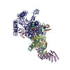 6n7rMC 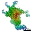 0360C 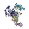 6n7pC C: citing same article ( M: atomic model generated by this map |
|---|---|
| Similar structure data |

-Links
| EMDB pages | EMDB (EBI/PDBe) / EMDataResource |
|---|---|
| Related items in Molecule of the Month |


-Map
| File | Download / File: emd_0361.map.gz / Format: CCP4 / Size: 166.4 MB / Type: IMAGE STORED AS FLOATING POINT NUMBER (4 BYTES) | ||||||||||||||||||||||||||||||||||||||||||||||||||||||||||||
|---|---|---|---|---|---|---|---|---|---|---|---|---|---|---|---|---|---|---|---|---|---|---|---|---|---|---|---|---|---|---|---|---|---|---|---|---|---|---|---|---|---|---|---|---|---|---|---|---|---|---|---|---|---|---|---|---|---|---|---|---|---|
| Annotation | Spliceosomal E complex (ACT1) | ||||||||||||||||||||||||||||||||||||||||||||||||||||||||||||
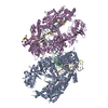
| Projections & slices | Image control
Images are generated by Spider. | ||||||||||||||||||||||||||||||||||||||||||||||||||||||||||||
| Voxel size | X=Y=Z: 1.36 Å | ||||||||||||||||||||||||||||||||||||||||||||||||||||||||||||
| Density |
| ||||||||||||||||||||||||||||||||||||||||||||||||||||||||||||
| Symmetry | Space group: 1 | ||||||||||||||||||||||||||||||||||||||||||||||||||||||||||||
| Details | EMDB XML:
CCP4 map header:
| ||||||||||||||||||||||||||||||||||||||||||||||||||||||||||||
 Z (Sec.)
 Y (Row.)
 X (Col.)


-Supplemental data
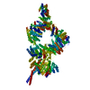
- Sample components
+Entire : Saccharomyces cerevisiae spliceosomal E complex (ACT1)
+Supramolecule #1: Saccharomyces cerevisiae spliceosomal E complex (ACT1)
+Macromolecule #1: U1 small nuclear ribonucleoprotein 70 kDa homolog
+Macromolecule #2: U1 small nuclear ribonucleoprotein C
+Macromolecule #3: U1 small nuclear ribonucleoprotein A
+Macromolecule #4: U1 small nuclear ribonucleoprotein component PRP42
+Macromolecule #5: Pre-mRNA-processing factor 39
+Macromolecule #6: Protein NAM8
+Macromolecule #7: 56 kDa U1 small nuclear ribonucleoprotein component
+Macromolecule #8: U1 small nuclear ribonucleoprotein component SNU71,U1 small nucle...
+Macromolecule #9: Protein LUC7
+Macromolecule #10: Small nuclear ribonucleoprotein-associated protein B
+Macromolecule #11: Small nuclear ribonucleoprotein Sm D1
+Macromolecule #12: Small nuclear ribonucleoprotein Sm D2
+Macromolecule #13: Small nuclear ribonucleoprotein Sm D3
+Macromolecule #14: Small nuclear ribonucleoprotein E
+Macromolecule #15: Small nuclear ribonucleoprotein F
+Macromolecule #16: Small nuclear ribonucleoprotein G
+Macromolecule #17: U1 snRNA
+Macromolecule #18: ACT1 pre-mRNA
+Macromolecule #19: ZINC ION
-Experimental details
-Structure determination
| Method | cryo EM |
|---|---|
 Processing Processing | single particle reconstruction |
| Aggregation state | particle |
-Sample preparation
| Buffer | pH: 7.9 |
|---|---|
| Grid | Details: unspecified |
| Vitrification | Cryogen name: ETHANE |
- Electron microscopy
| Microscope | FEI TITAN KRIOS |
|---|---|
| Image recording | Film or detector model: GATAN K2 SUMMIT (4k x 4k) / Detector mode: SUPER-RESOLUTION / Average electron dose: 30.0 e/Å2 |
| Electron beam | Acceleration voltage: 300 kV / Electron source:  FIELD EMISSION GUN FIELD EMISSION GUN |
| Electron optics | Illumination mode: FLOOD BEAM / Imaging mode: BRIGHT FIELD |
| Experimental equipment |  Model: Titan Krios / Image courtesy: FEI Company |

