 Movie
Movie Controller

[English] 日本語
 Yorodumi
- PDB-5xte: Cryo-EM structure of human respiratory complex III (cytochrome bc... -
+ Open data

- Basic information
| Entry | Database: PDB / ID: 5xte | ||||||||||||||||||
|---|---|---|---|---|---|---|---|---|---|---|---|---|---|---|---|---|---|---|---|
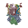
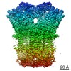
| Title | Cryo-EM structure of human respiratory complex III (cytochrome bc1 complex) | ||||||||||||||||||
 Components Components |
| ||||||||||||||||||
 Keywords Keywords | OXIDOREDUCTASE/ELECTRON TRANSPORT / Respiratory / OXIDOREDUCTASE-ELECTRON TRANSPORT complex | ||||||||||||||||||
| Function / homology |  Function and homology information Function and homology informationComplex III assembly / Complex III assembly / Complex III assembly / Complex III assembly / Complex III assembly / response to D-galactosamine / Complex III assembly / response to cobalamin / Complex III assembly / Complex III assembly ...Complex III assembly / Complex III assembly / Complex III assembly / Complex III assembly / Complex III assembly / response to D-galactosamine / Complex III assembly / response to cobalamin / Complex III assembly / Complex III assembly / Complex III assembly / response to mercury ion / subthalamus development / pons development / Respiratory electron transport / cerebellar Purkinje cell layer development / mitochondrial respiratory chain complex III assembly / pyramidal neuron development / thalamus development / response to alkaloid / Mitochondrial protein import / Mitochondrial translation termination / respiratory chain complex / respiratory chain complex III / cellular respiration / response to glucagon / oxidative phosphorylation / quinol-cytochrome-c reductase / quinol-cytochrome-c reductase activity / mitochondrial electron transport, ubiquinol to cytochrome c / response to copper ion / hypothalamus development / midbrain development / electron transport coupled proton transport / animal organ regeneration / response to hyperoxia / response to cadmium ion / Mitochondrial protein degradation / aerobic respiration / response to activity / respiratory electron transport chain / hippocampus development / generation of precursor metabolites and energy / metalloendopeptidase activity / response to calcium ion / 2 iron, 2 sulfur cluster binding / response to toxic substance / response to ethanol / response to hypoxia / electron transfer activity / oxidoreductase activity / mitochondrial inner membrane / mitochondrial matrix / response to xenobiotic stimulus / heme binding / ubiquitin protein ligase binding / protein-containing complex binding / mitochondrion / proteolysis / nucleoplasm / membrane / metal ion binding / nucleus Similarity search - Function | ||||||||||||||||||
| Biological species |  Homo sapiens (human) Homo sapiens (human) | ||||||||||||||||||
| Method | ELECTRON MICROSCOPY / single particle reconstruction / cryo EM / Resolution: 3.4 Å | ||||||||||||||||||
 Authors Authors | Gu, J. / Wu, M. / Yang, M. | ||||||||||||||||||
| Funding support |  China, 5items China, 5items
| ||||||||||||||||||
 Citation Citation | Journal: Cell / Year: 2017 Title: Architecture of Human Mitochondrial Respiratory Megacomplex IIIIIV. Authors: Runyu Guo / Shuai Zong / Meng Wu / Jinke Gu / Maojun Yang / Abstract: The respiratory megacomplex represents the highest-order assembly of respiratory chain complexes, and it allows mitochondria to respond to energy-requiring conditions. To understand its architecture, ...The respiratory megacomplex represents the highest-order assembly of respiratory chain complexes, and it allows mitochondria to respond to energy-requiring conditions. To understand its architecture, we examined the human respiratory chain megacomplex-IIIIIV (MCIIIIIV) with 140 subunits and a subset of associated cofactors using cryo-electron microscopy. The MCIIIIIV forms a circular structure with the dimeric CIII located in the center, where it is surrounded by two copies each of CI and CIV. Two cytochrome c (Cyt.c) molecules are positioned to accept electrons on the surface of the c state CIII dimer. Analyses indicate that CII could insert into the gaps between CI and CIV to form a closed ring, which we termed the electron transport chain supercomplex. The structure not only reveals the precise assignment of individual subunits of human CI and CIII, but also enables future in-depth analysis of the electron transport chain as a whole. | ||||||||||||||||||
| History |
|
- Structure visualization
| Movie |
Movie viewer |
|---|---|
| Structure viewer | Molecule: MolmilJmol/JSmol |
- Downloads & links
-Download
| PDBx/mmCIF format | 5xte.cif.gz | 852.3 KB | Display | PDBx/mmCIF format |
|---|---|---|---|---|
| PDB format | pdb5xte.ent.gz | 690.1 KB | Display | PDB format |
| PDBx/mmJSON format | 5xte.json.gz | Tree view | PDBx/mmJSON format | |
| Others |  Other downloads Other downloads |
-Validation report
| Arichive directory | https://data.pdbj.org/pub/pdb/validation_reports/xt/5xteftp://data.pdbj.org/pub/pdb/validation_reports/xt/5xte | HTTPS FTP |
|---|
-Related structure data
| Related structure data | 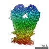 6774MC 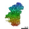 6771C 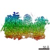 6772C 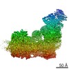 6773C 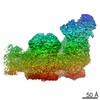 6775C 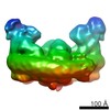 6776C 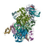 5xtbC 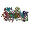 5xtcC  5xtdC  5xthC  5xtiC M: map data used to model this data C: citing same article ( |
|---|---|
| Similar structure data |


-Links
 PDBj
PDBj


- Assembly
| Deposited unit | 
|
|---|---|
| 1 |
|
-Components
-Cytochrome b-c1 complex subunit ... , 9 types, 18 molecules ANBOCPDQERFSGTKWLY
| #1: Protein | Mass: 9791.181 Da / Num. of mol.: 2 / Source method: isolated from a natural source / Source: (natural) Homo sapiens (human) / References: UniProt: O14949#2: Protein | Mass: 5893.814 Da / Num. of mol.: 2 / Fragment: UNP RESIDUES 1-57 / Source method: isolated from a natural source / Source: (natural) Homo sapiens (human) / References: UniProt: P47985, quinol-cytochrome-c reductase#3: Protein | Mass: 21645.578 Da / Num. of mol.: 2 / Source method: isolated from a natural source / Source: (natural) Homo sapiens (human) / References: UniProt: P47985, quinol-cytochrome-c reductase#4: Protein | Mass: 7189.299 Da / Num. of mol.: 2 / Source method: isolated from a natural source / Source: (natural) Homo sapiens (human) / References: UniProt: Q9UDW1#5: Protein | Mass: 8990.854 Da / Num. of mol.: 2 / Source method: isolated from a natural source / Source: (natural) Homo sapiens (human) / References: UniProt: P07919#6: Protein | Mass: 13037.842 Da / Num. of mol.: 2 / Fragment: UNP RESIDUES 6-111 / Source method: isolated from a natural source / Source: (natural) Homo sapiens (human) / References: UniProt: P14927#7: Protein | Mass: 5958.912 Da / Num. of mol.: 2 / Fragment: UNP RESIDUES 2-52 / Source method: isolated from a natural source / Source: (natural) Homo sapiens (human) / References: UniProt: O14957#10: Protein | Mass: 44946.582 Da / Num. of mol.: 2 / Fragment: UNP RESIDUES 35-453 / Source method: isolated from a natural source / Source: (natural) Homo sapiens (human) / References: UniProt: P22695#11: Protein | Mass: 49181.430 Da / Num. of mol.: 2 / Source method: isolated from a natural source / Source: (natural) Homo sapiens (human) / References: UniProt: P31930 |
|---|
-Protein , 2 types, 4 molecules HUJV
| #8: Protein | Mass: 27388.395 Da / Num. of mol.: 2 / Source method: isolated from a natural source / Source: (natural) Homo sapiens (human) / References: UniProt: P08574#9: Protein | Mass: 42543.016 Da / Num. of mol.: 2 / Source method: isolated from a natural source / Source: (natural) Homo sapiens (human) / References: UniProt: P00156 |
|---|
-Non-polymers , 6 types, 26 molecules 


| #12: Chemical | ChemComp-CDL /  Mass: 1464.043 Da / Num. of mol.: 9 / Source method: obtained synthetically / Formula: C81H156O17P2 / Comment: phospholipid*YM Mass: 1464.043 Da / Num. of mol.: 9 / Source method: obtained synthetically / Formula: C81H156O17P2 / Comment: phospholipid*YM#13: Chemical |  Mass: 175.820 Da / Num. of mol.: 2 / Source method: obtained synthetically / Formula: Fe2S2 Mass: 175.820 Da / Num. of mol.: 2 / Source method: obtained synthetically / Formula: Fe2S2#14: Chemical | ChemComp-PEE /  Mass: 744.034 Da / Num. of mol.: 6 / Source method: obtained synthetically / Formula: C41H78NO8P / Comment: DOPE, phospholipid*YM Mass: 744.034 Da / Num. of mol.: 6 / Source method: obtained synthetically / Formula: C41H78NO8P / Comment: DOPE, phospholipid*YM#15: Chemical |  Mass: 618.503 Da / Num. of mol.: 2 / Source method: obtained synthetically / Formula: C34H34FeN4O4 Mass: 618.503 Da / Num. of mol.: 2 / Source method: obtained synthetically / Formula: C34H34FeN4O4#16: Chemical | ChemComp-HEM /  Mass: 616.487 Da / Num. of mol.: 4 / Source method: obtained synthetically / Formula: C34H32FeN4O4 Mass: 616.487 Da / Num. of mol.: 4 / Source method: obtained synthetically / Formula: C34H32FeN4O4#17: Chemical |  Mass: 767.132 Da / Num. of mol.: 3 / Source method: obtained synthetically / Formula: C42H89NO8P / Comment: phospholipid*YM Mass: 767.132 Da / Num. of mol.: 3 / Source method: obtained synthetically / Formula: C42H89NO8P / Comment: phospholipid*YM |
|---|
-Details
| Has protein modification | Y |
|---|
-Experimental details
-Experiment
| Experiment | Method: ELECTRON MICROSCOPY |
|---|---|
| EM experiment | Aggregation state: PARTICLE / 3D reconstruction method: single particle reconstruction |
- Sample preparation
| Component | Name: Human respiratory complex III (cytochrome bc1 complex) Type: COMPLEX / Entity ID: #1-#11 / Source: NATURAL |
|---|---|
| Source (natural) | Organism: Homo sapiens (human) |
| Buffer solution | pH: 7.4 |
| Specimen | Embedding applied: NO / Shadowing applied: NO / Staining applied: NO / Vitrification applied: YES |
| Vitrification | Cryogen name: ETHANE |
- Electron microscopy imaging
| Experimental equipment |  Model: Titan Krios / Image courtesy: FEI Company |
|---|---|
| Microscopy | Model: FEI TITAN KRIOS |
| Electron gun | Electron source:  FIELD EMISSION GUN / Accelerating voltage: 300 kV / Illumination mode: OTHER FIELD EMISSION GUN / Accelerating voltage: 300 kV / Illumination mode: OTHER |
| Electron lens | Mode: BRIGHT FIELD |
| Image recording | Electron dose: 1.25 e/Å2 / Film or detector model: FEI FALCON II (4k x 4k) |
- Processing
| EM software |
| ||||||||||||||||||||||||||||||||||||||||
|---|---|---|---|---|---|---|---|---|---|---|---|---|---|---|---|---|---|---|---|---|---|---|---|---|---|---|---|---|---|---|---|---|---|---|---|---|---|---|---|---|---|
| CTF correction | Type: NONE | ||||||||||||||||||||||||||||||||||||||||
| Symmetry | Point symmetry: C1 (asymmetric) | ||||||||||||||||||||||||||||||||||||||||
| 3D reconstruction | Resolution: 3.4 Å / Resolution method: FSC 0.143 CUT-OFF / Num. of particles: 167761 / Symmetry type: POINT |
