 Movie
Movie Controller

[English] 日本語
 Yorodumi
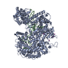
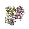
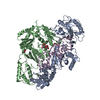
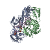
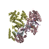
- PDB-5wlh: Crystal structure of LbaCas13a H328A (C2c2) bound to pre-crRNA (2... -
+ Open data

- Basic information
| Entry | Database: PDB / ID: 5wlh | ||||||
|---|---|---|---|---|---|---|---|
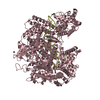
| Title | Crystal structure of LbaCas13a H328A (C2c2) bound to pre-crRNA (24-nt spacer) | ||||||
 Components Components |
| ||||||
 Keywords Keywords | RNA BINDING PROTEIN/RNA / Nuclease RNA binding protein / RNA BINDING PROTEIN / RNA BINDING PROTEIN-RNA complex | ||||||
| Function / homology | : / nuclease activity / defense response to virus / RNA binding / IODIDE ION / RNA / RNA (> 10) / CRISPR-associated endoribonuclease Cas13a Function and homology information Function and homology information | ||||||
| Biological species |  Lachnospiraceae bacterium NK4A179 (bacteria) Lachnospiraceae bacterium NK4A179 (bacteria) | ||||||
| Method |  X-RAY DIFFRACTION / SYNCHROTRON / MOLECULAR REPLACEMENT / Resolution: 1.80001033182 Å X-RAY DIFFRACTION / SYNCHROTRON / MOLECULAR REPLACEMENT / Resolution: 1.80001033182 Å | ||||||
 Authors Authors | Knott, G.J. / Doudna, J.A. | ||||||
 Citation Citation | Journal: Nat. Struct. Mol. Biol. / Year: 2017 Title: Guide-bound structures of an RNA-targeting A-cleaving CRISPR-Cas13a enzyme. Authors: Knott, G.J. / East-Seletsky, A. / Cofsky, J.C. / Holton, J.M. / Charles, E. / O'Connell, M.R. / Doudna, J.A. | ||||||
| History |
|
- Structure visualization
| Structure viewer | Molecule: MolmilJmol/JSmol |
|---|
- Downloads & links
-Download
| PDBx/mmCIF format | 5wlh.cif.gz | 398.8 KB | Display | PDBx/mmCIF format |
|---|---|---|---|---|
| PDB format | pdb5wlh.ent.gz | 251 KB | Display | PDB format |
| PDBx/mmJSON format | 5wlh.json.gz | Tree view | PDBx/mmJSON format | |
| Others |  Other downloads Other downloads |
-Validation report
| Arichive directory | https://data.pdbj.org/pub/pdb/validation_reports/wl/5wlhftp://data.pdbj.org/pub/pdb/validation_reports/wl/5wlh | HTTPS FTP |
|---|
-Related structure data
| Related structure data | 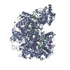 5w1hC  5w1iC 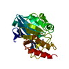 5wihS C: citing same article ( S: Starting model for refinement |
|---|---|
| Similar structure data |


-Links
 PDBj
PDBj


- Assembly
| Deposited unit | 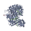
| ||||||||||||
|---|---|---|---|---|---|---|---|---|---|---|---|---|---|
| 1 |
| ||||||||||||
| Unit cell |
|
-Components
| #1: Protein | Mass: 169125.641 Da / Num. of mol.: 1 / Mutation: H328A Source method: isolated from a genetically manipulated source Source: (gene. exp.) Lachnospiraceae bacterium NK4A179 (bacteria)Production host: | ||||
|---|---|---|---|---|---|
| #2: RNA chain | Mass: 17563.594 Da / Num. of mol.: 1 / Source method: obtained synthetically Source: (synth.) Lachnospiraceae bacterium NK4A179 (bacteria) | ||||
| #3: Chemical | ChemComp-IOD /   Mass: 126.904 Da / Num. of mol.: 20 / Source method: obtained synthetically / Formula: I Mass: 126.904 Da / Num. of mol.: 20 / Source method: obtained synthetically / Formula: I#4: Chemical | ChemComp-GOL / |   Mass: 92.094 Da / Num. of mol.: 1 / Source method: obtained synthetically / Formula: C3H8O3 Mass: 92.094 Da / Num. of mol.: 1 / Source method: obtained synthetically / Formula: C3H8O3#5: Water | ChemComp-HOH / |  Mass: 18.015 Da / Num. of mol.: 765 / Source method: isolated from a natural source / Formula: H2O Mass: 18.015 Da / Num. of mol.: 765 / Source method: isolated from a natural source / Formula: H2O |
-Experimental details
-Experiment
| Experiment | Method: X-RAY DIFFRACTION / Number of used crystals: 1 |
|---|
- Sample preparation
| Crystal | Density Matthews: 2.16 Å3/Da / Density % sol: 42.95 % |
|---|---|
| Crystal grow | Temperature: 293 K / Method: vapor diffusion, sitting drop / pH: 7 / Details: 0.2 M ammonium iodide, 20 % PEG 3,350 |
-Data collection
| Diffraction | Mean temperature: 100 K | ||||||||||||||||||||||||||||||
|---|---|---|---|---|---|---|---|---|---|---|---|---|---|---|---|---|---|---|---|---|---|---|---|---|---|---|---|---|---|---|---|
| Diffraction source | Source: SYNCHROTRON / Site: ALS  / Beamline: 8.3.1 / Wavelength: 1.1158 Å / Beamline: 8.3.1 / Wavelength: 1.1158 Å | ||||||||||||||||||||||||||||||
| Detector | Type: DECTRIS PILATUS3 6M / Detector: PIXEL / Date: Jul 18, 2017 | ||||||||||||||||||||||||||||||
| Radiation | Protocol: SINGLE WAVELENGTH / Monochromatic (M) / Laue (L): M / Scattering type: x-ray | ||||||||||||||||||||||||||||||
| Radiation wavelength | Wavelength: 1.1158 Å / Relative weight: 1 | ||||||||||||||||||||||||||||||
| Reflection | Resolution: 1.71→47.116494566 Å / Num. obs: 162397 / % possible obs: 93.3 % / Redundancy: 5 % / Biso Wilson estimate: 30.1855803653 Å2 / CC1/2: 0.999 / Rmerge(I) obs: 0.051 / Rpim(I) all: 0.025 / Rrim(I) all: 0.057 / Χ2: 1.18 / Net I/σ(I): 15.2 / Num. measured all: 807018 / Scaling rejects: 4 | ||||||||||||||||||||||||||||||
| Reflection shell | Diffraction-ID: 1
|
- Processing
| Software |
| |||||||||||||||||||||||||||||||||||||||||||||||||||||||||||||||||||||||||||||||||||||||||||||||||||||||||||||||||||||||||||||||||||||||||||||||||||||||||||||||||||||||||||||||||||||||||||||||||||||||||||||||||||||||||
|---|---|---|---|---|---|---|---|---|---|---|---|---|---|---|---|---|---|---|---|---|---|---|---|---|---|---|---|---|---|---|---|---|---|---|---|---|---|---|---|---|---|---|---|---|---|---|---|---|---|---|---|---|---|---|---|---|---|---|---|---|---|---|---|---|---|---|---|---|---|---|---|---|---|---|---|---|---|---|---|---|---|---|---|---|---|---|---|---|---|---|---|---|---|---|---|---|---|---|---|---|---|---|---|---|---|---|---|---|---|---|---|---|---|---|---|---|---|---|---|---|---|---|---|---|---|---|---|---|---|---|---|---|---|---|---|---|---|---|---|---|---|---|---|---|---|---|---|---|---|---|---|---|---|---|---|---|---|---|---|---|---|---|---|---|---|---|---|---|---|---|---|---|---|---|---|---|---|---|---|---|---|---|---|---|---|---|---|---|---|---|---|---|---|---|---|---|---|---|---|---|---|---|---|---|---|---|---|---|---|---|---|---|---|---|---|---|---|---|
| Refinement | Method to determine structure: MOLECULAR REPLACEMENT Starting model: 5WIH Resolution: 1.80001033182→47.116494566 Å / SU ML: 0.215930613219 / Cross valid method: FREE R-VALUE / σ(F): 1.35143778714 / Phase error: 23.5449843848 Stereochemistry target values: GeoStd + Monomer Library + CDL v1.2
| |||||||||||||||||||||||||||||||||||||||||||||||||||||||||||||||||||||||||||||||||||||||||||||||||||||||||||||||||||||||||||||||||||||||||||||||||||||||||||||||||||||||||||||||||||||||||||||||||||||||||||||||||||||||||
| Solvent computation | Shrinkage radii: 0.9 Å / VDW probe radii: 1.11 Å / Solvent model: FLAT BULK SOLVENT MODEL | |||||||||||||||||||||||||||||||||||||||||||||||||||||||||||||||||||||||||||||||||||||||||||||||||||||||||||||||||||||||||||||||||||||||||||||||||||||||||||||||||||||||||||||||||||||||||||||||||||||||||||||||||||||||||
| Displacement parameters | Biso mean: 37.3534789683 Å2 | |||||||||||||||||||||||||||||||||||||||||||||||||||||||||||||||||||||||||||||||||||||||||||||||||||||||||||||||||||||||||||||||||||||||||||||||||||||||||||||||||||||||||||||||||||||||||||||||||||||||||||||||||||||||||
| Refinement step | Cycle: LAST / Resolution: 1.80001033182→47.116494566 Å
| |||||||||||||||||||||||||||||||||||||||||||||||||||||||||||||||||||||||||||||||||||||||||||||||||||||||||||||||||||||||||||||||||||||||||||||||||||||||||||||||||||||||||||||||||||||||||||||||||||||||||||||||||||||||||
| Refine LS restraints |
| |||||||||||||||||||||||||||||||||||||||||||||||||||||||||||||||||||||||||||||||||||||||||||||||||||||||||||||||||||||||||||||||||||||||||||||||||||||||||||||||||||||||||||||||||||||||||||||||||||||||||||||||||||||||||
| LS refinement shell |
|
