 Movie
Movie Controller

[English] 日本語
 Yorodumi
- PDB-3v4i: Crystal structure of HIV-1 reverse transcriptase (RT) with DNA an... -
+ Open data

- Basic information
| Entry | Database: PDB / ID: 3v4i | ||||||
|---|---|---|---|---|---|---|---|
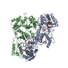
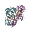
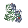
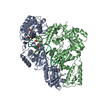
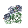
| Title | Crystal structure of HIV-1 reverse transcriptase (RT) with DNA and AZTTP | ||||||
 Components Components |
| ||||||
 Keywords Keywords | TRANSFERASE/DNA / HIV-1 reverse transcriptase / zidovudine / RT-DNA complex / transferase-dna complex / drug resistance mutation / AIDS / DNA recombination / DNA-directed DNA polymerase / RNASE H / hydrolase / lipoprotein / magnesium / membrane / metal-binding / multifunctional enzyme / nucleotidyltransferase / RNA-directed DNA polymerase transferase / transferase-DNA complex complex | ||||||
| Function / homology |  Function and homology information Function and homology informationHIV-1 retropepsin / symbiont-mediated activation of host apoptosis / retroviral ribonuclease H / exoribonuclease H / exoribonuclease H activity / DNA integration / viral genome integration into host DNA / establishment of integrated proviral latency / RNA-directed DNA polymerase / RNA stem-loop binding ...HIV-1 retropepsin / symbiont-mediated activation of host apoptosis / retroviral ribonuclease H / exoribonuclease H / exoribonuclease H activity / DNA integration / viral genome integration into host DNA / establishment of integrated proviral latency / RNA-directed DNA polymerase / RNA stem-loop binding / viral penetration into host nucleus / host multivesicular body / RNA-directed DNA polymerase activity / RNA-DNA hybrid ribonuclease activity / Transferases; Transferring phosphorus-containing groups; Nucleotidyltransferases / host cell / viral nucleocapsid / DNA recombination / DNA-directed DNA polymerase / aspartic-type endopeptidase activity / Hydrolases; Acting on ester bonds / DNA-directed DNA polymerase activity / symbiont-mediated suppression of host gene expression / viral translational frameshifting / symbiont entry into host cell / lipid binding / host cell nucleus / host cell plasma membrane / virion membrane / structural molecule activity / proteolysis / DNA binding / zinc ion binding Similarity search - Function | ||||||
| Biological species |  Human immunodeficiency virus type 1 BH10 Human immunodeficiency virus type 1 BH10 | ||||||
| Method |  X-RAY DIFFRACTION / SYNCHROTRON / MOLECULAR REPLACEMENT / Resolution: 2.7983 Å X-RAY DIFFRACTION / SYNCHROTRON / MOLECULAR REPLACEMENT / Resolution: 2.7983 Å | ||||||
 Authors Authors | Das, K. / Martinez, S.E. / Arnold, E. | ||||||
 Citation Citation | Journal: Nat.Struct.Mol.Biol. / Year: 2012 Title: HIV-1 reverse transcriptase complex with DNA and nevirapine reveals non-nucleoside inhibition mechanism. Authors: Das, K. / Martinez, S.E. / Bauman, J.D. / Arnold, E. #1: Journal: J.Biol.Chem. / Year: 2009Title: Structural basis for the role of the K65R mutation in HIV-1 reverse transcriptase polymerization, excision antagonism, and tenofovir resistance. Authors: Das, K. / Bandwar, R.P. / White, K.L. / Feng, J.Y. / Sarafianos, S.G. / Tuske, S. / Tu, X. / Clark, A.D. / Boyer, P.L. / Hou, X. / Gaffney, B.L. / Jones, R.A. / Miller, M.D. / Hughes, S.H. / Arnold, E. #2: Journal: Nat.Struct.Mol.Biol. / Year: 2010Title: Structural basis of HIV-1 resistance to AZT by excision. Authors: Tu, X. / Das, K. / Han, Q. / Bauman, J.D. / Clark, A.D. / Hou, X. / Frenkel, Y.V. / Gaffney, B.L. / Jones, R.A. / Boyer, P.L. / Hughes, S.H. / Sarafianos, S.G. / Arnold, E. #3: Journal: Proc.Natl.Acad.Sci.USA / Year: 2008Title: High-resolution structures of HIV-1 reverse transcriptase/TMC278 complexes: strategic flexibility explains potency against resistance mutations. Authors: Das, K. / Bauman, J.D. / Clark, A.D. / Frenkel, Y.V. / Lewi, P.J. / Shatkin, A.J. / Hughes, S.H. / Arnold, E. #4: Journal: J.Med.Chem. / Year: 2004Title: Roles of conformational and positional adaptability in structure-based design of TMC125-R165335 (etravirine) and related non-nucleoside reverse transcriptase inhibitors that are highly potent ...Title: Roles of conformational and positional adaptability in structure-based design of TMC125-R165335 (etravirine) and related non-nucleoside reverse transcriptase inhibitors that are highly potent and effective against wild-type and drug-resistant HIV-1 variants. Authors: Das, K. / Clark, A.D. / Lewi, P.J. / Heeres, J. / De Jonge, M.R. / Koymans, L.M. / Vinkers, H.M. / Daeyaert, F. / Ludovici, D.W. / Kukla, M.J. / De Corte, B. / Kavash, R.W. / Ho, C.Y. / Ye, ...Authors: Das, K. / Clark, A.D. / Lewi, P.J. / Heeres, J. / De Jonge, M.R. / Koymans, L.M. / Vinkers, H.M. / Daeyaert, F. / Ludovici, D.W. / Kukla, M.J. / De Corte, B. / Kavash, R.W. / Ho, C.Y. / Ye, H. / Lichtenstein, M.A. / Andries, K. / Pauwels, R. / De Bethune, M.P. / Boyer, P.L. / Clark, P. / Hughes, S.H. / Janssen, P.A. / Arnold, E. #5: Journal: Science / Year: 1998Title: Structure of a covalently trapped catalytic complex of HIV-1 reverse transcriptase: implications for drug resistance. Authors: Huang, H. / Chopra, R. / Verdine, G.L. / Harrison, S.C. | ||||||
| History |
|
- Structure visualization
| Structure viewer | Molecule: MolmilJmol/JSmol |
|---|
- Downloads & links
-Download
| PDBx/mmCIF format | 3v4i.cif.gz | 456.3 KB | Display | PDBx/mmCIF format |
|---|---|---|---|---|
| PDB format | pdb3v4i.ent.gz | 361.9 KB | Display | PDB format |
| PDBx/mmJSON format | 3v4i.json.gz | Tree view | PDBx/mmJSON format | |
| Others |  Other downloads Other downloads |
-Validation report
| Arichive directory | https://data.pdbj.org/pub/pdb/validation_reports/v4/3v4iftp://data.pdbj.org/pub/pdb/validation_reports/v4/3v4i | HTTPS FTP |
|---|
-Related structure data
| Related structure data | 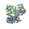 3v6dC  3v81C 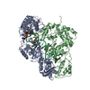 3jsmS C: citing same article ( S: Starting model for refinement |
|---|---|
| Similar structure data |


-Links
 PDBj
PDBj


- Assembly
| Deposited unit | 
| ||||||||
|---|---|---|---|---|---|---|---|---|---|
| 1 | 
| ||||||||
| 2 | 
| ||||||||
| Unit cell |
|
-Components
-HIV-1 Reverse Transcriptase ... , 2 types, 4 molecules ACBD
| #1: Protein | Mass: 64022.414 Da / Num. of mol.: 2 / Fragment: UNP Residues 600-1153 / Mutation: D498N Source method: isolated from a genetically manipulated source Source: (gene. exp.) Human immunodeficiency virus type 1 BH10Gene: gag-pol / Production host:  References: UniProt: P03366, RNA-directed DNA polymerase, DNA-directed DNA polymerase #2: Protein | Mass: 50039.488 Da / Num. of mol.: 2 / Fragment: UNP Residues 600-1027 Source method: isolated from a genetically manipulated source Source: (gene. exp.) Human immunodeficiency virus type 1 BH10Gene: gag-pol / Production host: References: UniProt: P03366, RNA-directed DNA polymerase, DNA-directed DNA polymerase |
|---|
-DNA chain , 2 types, 4 molecules TEPF
| #3: DNA chain | Mass: 8416.424 Da / Num. of mol.: 2 / Source method: obtained synthetically / Details: Synthesized #4: DNA chain | Mass: 6490.267 Da / Num. of mol.: 2 / Source method: obtained synthetically / Details: Synthesized |
|---|
-Non-polymers , 2 types, 4 molecules 

| #5: Chemical |  Mass: 24.305 Da / Num. of mol.: 2 / Source method: obtained synthetically / Formula: Mg Mass: 24.305 Da / Num. of mol.: 2 / Source method: obtained synthetically / Formula: Mg#6: Chemical |  Mass: 507.181 Da / Num. of mol.: 2 / Source method: obtained synthetically / Formula: C10H16N5O13P3 Mass: 507.181 Da / Num. of mol.: 2 / Source method: obtained synthetically / Formula: C10H16N5O13P3 |
|---|
-Details
| Has protein modification | Y |
|---|
-Experimental details
-Experiment
| Experiment | Method: X-RAY DIFFRACTION / Number of used crystals: 1 |
|---|
- Sample preparation
| Crystal | Density Matthews: 3.19 Å3/Da / Density % sol: 61.48 % |
|---|---|
| Crystal grow | Temperature: 277 K / Method: evaporation / pH: 6.8 Details: PEG 8000, AMMONIUM SULFATE, MGCL2, GLYCEROL, SUCROSE, pH 7.2, EVAPORATION, temperature 277K |
-Data collection
| Diffraction | Mean temperature: 100 K |
|---|---|
| Diffraction source | Source: SYNCHROTRON / Site: CHESS  / Beamline: F1 / Wavelength: 0.918 Å / Beamline: F1 / Wavelength: 0.918 Å |
| Detector | Type: ADSC QUANTUM 270 / Detector: CCD / Date: Dec 1, 2010 |
| Radiation | Protocol: SINGLE WAVELENGTH / Monochromatic (M) / Laue (L): M / Scattering type: x-ray |
| Radiation wavelength | Wavelength: 0.918 Å / Relative weight: 1 |
| Reflection | Resolution: 2.798→50 Å / Num. obs: 73202 / % possible obs: 92.8 % / Observed criterion σ(F): 0 / Observed criterion σ(I): -1 / Redundancy: 3.2 % / Rmerge(I) obs: 0.083 / Net I/σ(I): 11.6 |
| Reflection shell | Resolution: 2.798→2.9 Å / Redundancy: 2.4 % / Rmerge(I) obs: 0.523 / Mean I/σ(I) obs: 1.9 / Num. unique all: 6702 / % possible all: 85.1 |
- Processing
| Software |
| |||||||||||||||||||||||||||||||||||||||||||||||||||||||||||||||||||||||||||||
|---|---|---|---|---|---|---|---|---|---|---|---|---|---|---|---|---|---|---|---|---|---|---|---|---|---|---|---|---|---|---|---|---|---|---|---|---|---|---|---|---|---|---|---|---|---|---|---|---|---|---|---|---|---|---|---|---|---|---|---|---|---|---|---|---|---|---|---|---|---|---|---|---|---|---|---|---|---|---|
| Refinement | Method to determine structure: MOLECULAR REPLACEMENT Starting model: PDB ENTRY 3JSM Resolution: 2.7983→45.543 Å / Occupancy max: 1 / Occupancy min: 1 / FOM work R set: 0.7767 / SU ML: 0.98 / σ(F): 0 / Phase error: 29.62 / Stereochemistry target values: Engh & Huber
| |||||||||||||||||||||||||||||||||||||||||||||||||||||||||||||||||||||||||||||
| Solvent computation | Shrinkage radii: 0.95 Å / VDW probe radii: 1.2 Å / Solvent model: FLAT BULK SOLVENT MODEL / Bsol: 27.18 Å2 / ksol: 0.302 e/Å3 | |||||||||||||||||||||||||||||||||||||||||||||||||||||||||||||||||||||||||||||
| Displacement parameters | Biso max: 169.4 Å2 / Biso mean: 63.8603 Å2 / Biso min: 16.89 Å2
| |||||||||||||||||||||||||||||||||||||||||||||||||||||||||||||||||||||||||||||
| Refinement step | Cycle: LAST / Resolution: 2.7983→45.543 Å
| |||||||||||||||||||||||||||||||||||||||||||||||||||||||||||||||||||||||||||||
| Refine LS restraints |
| |||||||||||||||||||||||||||||||||||||||||||||||||||||||||||||||||||||||||||||
| LS refinement shell | Refine-ID: X-RAY DIFFRACTION / Total num. of bins used: 10
|
