 Movie
Movie Controller

[English] 日本語
 Yorodumi
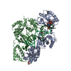
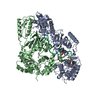
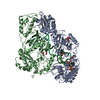
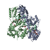
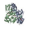
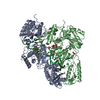
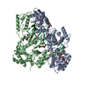
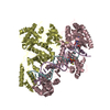
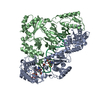
- PDB-3klf: Crystal structure of wild-type HIV-1 Reverse Transcriptase crossl... -
+ Open data

- Basic information
| Entry | Database: PDB / ID: 3klf | ||||||
|---|---|---|---|---|---|---|---|
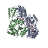
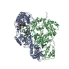
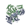
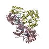
| Title | Crystal structure of wild-type HIV-1 Reverse Transcriptase crosslinked to a DSDNA with a bound excision product, AZTPPPPA | ||||||
 Components Components |
| ||||||
 Keywords Keywords | TRANSFERASE/DNA / AZT RESISTANCE MECHANISM / HIV-1 REVERSE TRANSCRIPTASE / WILD-TYPE / AZT RESISTANCE MUTATIONS / P51/P66 / NUCELEOSIDE INHIBITOR / THYMIDINE ANALOG MUTATIONS / AIDS / HIV / DNA POLYMERASE / NRTI / NRTI RESISTANCE / AZT / AZTPPPPA / AZTP4A / DINUCLEOSIDE TETRAPHOSPHATE / DNA-DIRECTED DNA POLYMERASE / ENDONUCLEASE / HYDROLASE / MAGNESIUM / METAL-BINDING / MULTIFUNCTIONAL ENZYME / NUCLEASE / RNA-DIRECTED DNA POLYMERASE / TRANSFERASE / TRANSFERASE-DNA COMPLEX | ||||||
| Function / homology |  Function and homology information Function and homology informationHIV-1 retropepsin / symbiont-mediated activation of host apoptosis / retroviral ribonuclease H / exoribonuclease H / exoribonuclease H activity / DNA integration / viral genome integration into host DNA / establishment of integrated proviral latency / RNA-directed DNA polymerase / RNA stem-loop binding ...HIV-1 retropepsin / symbiont-mediated activation of host apoptosis / retroviral ribonuclease H / exoribonuclease H / exoribonuclease H activity / DNA integration / viral genome integration into host DNA / establishment of integrated proviral latency / RNA-directed DNA polymerase / RNA stem-loop binding / viral penetration into host nucleus / host multivesicular body / RNA-directed DNA polymerase activity / RNA-DNA hybrid ribonuclease activity / Transferases; Transferring phosphorus-containing groups; Nucleotidyltransferases / host cell / viral nucleocapsid / DNA recombination / DNA-directed DNA polymerase / aspartic-type endopeptidase activity / Hydrolases; Acting on ester bonds / DNA-directed DNA polymerase activity / symbiont-mediated suppression of host gene expression / viral translational frameshifting / symbiont entry into host cell / lipid binding / host cell nucleus / host cell plasma membrane / virion membrane / structural molecule activity / proteolysis / DNA binding / zinc ion binding Similarity search - Function | ||||||
| Biological species |   Human immunodeficiency virus type 1 Human immunodeficiency virus type 1 | ||||||
| Method |  X-RAY DIFFRACTION / SYNCHROTRON / MOLECULAR REPLACEMENT / Resolution: 3.15 Å X-RAY DIFFRACTION / SYNCHROTRON / MOLECULAR REPLACEMENT / Resolution: 3.15 Å | ||||||
 Authors Authors | Tu, X. / Das, K. / Sarafianos, S.G. / Arnold, E. | ||||||
 Citation Citation | Journal: Nat.Struct.Mol.Biol. / Year: 2010 Title: Structural basis of HIV-1 resistance to AZT by excision. Authors: Tu, X. / Das, K. / Han, Q. / Bauman, J.D. / Clark, A.D. / Hou, X. / Frenkel, Y.V. / Gaffney, B.L. / Jones, R.A. / Boyer, P.L. / Hughes, S.H. / Sarafianos, S.G. / Arnold, E. | ||||||
| History |
|
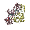
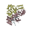
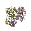
- Structure visualization
| Structure viewer | Molecule: MolmilJmol/JSmol |
|---|
- Downloads & links
-Download
| PDBx/mmCIF format | 3klf.cif.gz | 883.9 KB | Display | PDBx/mmCIF format |
|---|---|---|---|---|
| PDB format | pdb3klf.ent.gz | 710.2 KB | Display | PDB format |
| PDBx/mmJSON format | 3klf.json.gz | Tree view | PDBx/mmJSON format | |
| Others |  Other downloads Other downloads |
-Validation report
| Arichive directory | https://data.pdbj.org/pub/pdb/validation_reports/kl/3klfftp://data.pdbj.org/pub/pdb/validation_reports/kl/3klf | HTTPS FTP |
|---|
-Related structure data
| Related structure data | 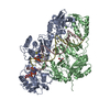 3kleC  3klgC  3klhC 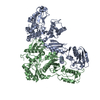 3kliC 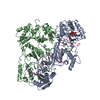 1rtdS C: citing same article ( S: Starting model for refinement |
|---|---|
| Similar structure data |


-Links
 PDBj
PDBj


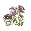
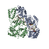
- Assembly
| Deposited unit | 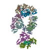
| ||||||||
|---|---|---|---|---|---|---|---|---|---|
| 1 | 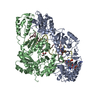
| ||||||||
| 2 | 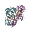
| ||||||||
| 3 | 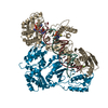
| ||||||||
| 4 | 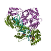
| ||||||||
| Unit cell |
|
-Components
-Protein , 2 types, 8 molecules AEIMBFJN
| #1: Protein | Mass: 64080.457 Da / Num. of mol.: 4 / Mutation: C280S, Q258C Source method: isolated from a genetically manipulated source Source: (gene. exp.) Human immunodeficiency virus type 1 / Strain: BH10 / Gene: gag-pol / Plasmid: PRT35A / Production host:  References: UniProt: P03366, RNA-directed DNA polymerase, DNA-directed DNA polymerase, ribonuclease H #2: Protein | Mass: 51928.629 Da / Num. of mol.: 4 / Mutation: C280S Source method: isolated from a genetically manipulated source Source: (gene. exp.) Human immunodeficiency virus type 1 / Strain: BH10 / Gene: gag-pol / Plasmid: PRT35A / Production host: |
|---|
-DNA chain , 2 types, 8 molecules CGKODHLP
| #3: DNA chain | Mass: 8367.386 Da / Num. of mol.: 4 / Source method: obtained synthetically / Details: DNA TEMPLATE FOR REVERSE TRANSCRIPTASE #4: DNA chain | Mass: 6458.268 Da / Num. of mol.: 4 / Source method: obtained synthetically / Details: DNA PRIMER FOR REVERSE TRANSCRIPTASE |
|---|
-Non-polymers , 4 types, 90 molecules 
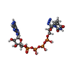


| #5: Chemical | ChemComp-MG /  Mass: 24.305 Da / Num. of mol.: 8 / Source method: obtained synthetically / Formula: Mg Mass: 24.305 Da / Num. of mol.: 8 / Source method: obtained synthetically / Formula: Mg#6: Chemical | ChemComp-ZP4 / [[[[(  Mass: 836.387 Da / Num. of mol.: 4 / Source method: obtained synthetically / Formula: C20H28N10O19P4 Mass: 836.387 Da / Num. of mol.: 4 / Source method: obtained synthetically / Formula: C20H28N10O19P4#7: Chemical |  Mass: 92.094 Da / Num. of mol.: 2 / Source method: obtained synthetically / Formula: C3H8O3 Mass: 92.094 Da / Num. of mol.: 2 / Source method: obtained synthetically / Formula: C3H8O3#8: Water | ChemComp-HOH / | Mass: 18.015 Da / Num. of mol.: 76 / Source method: isolated from a natural source / Formula: H2O |
|---|
-Details
| Has protein modification | Y |
|---|
-Experimental details
-Experiment
| Experiment | Method: X-RAY DIFFRACTION / Number of used crystals: 1 |
|---|
- Sample preparation
| Crystal | Density Matthews: 3.14 Å3/Da / Density % sol: 60.85 % |
|---|---|
| Crystal grow | pH: 6.8 Details: 50 MM BIS-TRIS PROPANE PH6.4, 10-11% PEG8000, 0.3 M (NH4)2SO4, 5% GLYCEROL, 5% SUCROSE, 20 MM MGCL2, AND 5 MM SPERMIDINE, PH 6.8, VAPOR DIFFUSION, HANGING DROP, TEMPERATURE 277.0K |
-Data collection
| Diffraction source | Source: SYNCHROTRON / Site: CHESS  / Beamline: A1 / Beamline: A1 |
|---|---|
| Detector | Type: ADSC QUANTUM 210 / Detector: CCD / Date: Feb 22, 2006 |
| Radiation | Protocol: SINGLE WAVELENGTH / Monochromatic (M) / Laue (L): M / Scattering type: x-ray |
| Radiation wavelength | Relative weight: 1 |
| Reflection | Resolution: 3.15→25 Å / Num. obs: 98589 / % possible obs: 90.3 % / Redundancy: 5.3 % / Rmerge(I) obs: 0.129 |
| Reflection shell | Resolution: 3.15→3.21 Å / Redundancy: 4.6 % / Rmerge(I) obs: 0.539 / % possible all: 83.5 |
- Processing
| Software |
| ||||||||||||||||||||||||||||||||||||||||||||||||||||||||||||
|---|---|---|---|---|---|---|---|---|---|---|---|---|---|---|---|---|---|---|---|---|---|---|---|---|---|---|---|---|---|---|---|---|---|---|---|---|---|---|---|---|---|---|---|---|---|---|---|---|---|---|---|---|---|---|---|---|---|---|---|---|---|
| Refinement | Method to determine structure: MOLECULAR REPLACEMENT Starting model: PDB ENTRY 1RTD Resolution: 3.15→24.83 Å / Rfactor Rfree error: 0.006 / Data cutoff high absF: 4028470 / Data cutoff low absF: 0 / Isotropic thermal model: OVERALL / Cross valid method: THROUGHOUT / σ(F): 0 / Stereochemistry target values: ENGH & HUBER / Details: BULK SOLVENT MODEL USED
| ||||||||||||||||||||||||||||||||||||||||||||||||||||||||||||
| Solvent computation | Solvent model: FLAT MODEL / Bsol: 41.82 Å2 / ksol: 0.3 e/Å3 | ||||||||||||||||||||||||||||||||||||||||||||||||||||||||||||
| Displacement parameters | Biso mean: 72.7 Å2
| ||||||||||||||||||||||||||||||||||||||||||||||||||||||||||||
| Refine analyze |
| ||||||||||||||||||||||||||||||||||||||||||||||||||||||||||||
| Refinement step | Cycle: LAST / Resolution: 3.15→24.83 Å
| ||||||||||||||||||||||||||||||||||||||||||||||||||||||||||||
| Refine LS restraints |
| ||||||||||||||||||||||||||||||||||||||||||||||||||||||||||||
| LS refinement shell | Resolution: 3.15→3.35 Å / Rfactor Rfree error: 0.023 / Total num. of bins used: 6
| ||||||||||||||||||||||||||||||||||||||||||||||||||||||||||||
| Xplor file |
|
