 Movie
Movie Controller

[English] 日本語
 Yorodumi
- PDB-6hak: Crystal structure of HIV-1 reverse transcriptase (RT) in complex ... -
+ Open data

- Basic information
| Entry | Database: PDB / ID: 6hak | |||||||||
|---|---|---|---|---|---|---|---|---|---|---|
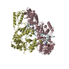
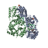
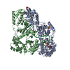
| Title | Crystal structure of HIV-1 reverse transcriptase (RT) in complex with a double stranded RNA represents the RT transcription initiation complex prior to nucleotide incorporation | |||||||||
 Components Components |
| |||||||||
 Keywords Keywords | TRANSCRIPTION / tRNA-Lys3 / Protein-RNA cross-link / transcription initiation / viral RNA / DNA polymerase | |||||||||
| Function / homology |  Function and homology information Function and homology informationHIV-1 retropepsin / symbiont-mediated activation of host apoptosis / retroviral ribonuclease H / exoribonuclease H / exoribonuclease H activity / DNA integration / viral genome integration into host DNA / establishment of integrated proviral latency / RNA-directed DNA polymerase / RNA stem-loop binding ...HIV-1 retropepsin / symbiont-mediated activation of host apoptosis / retroviral ribonuclease H / exoribonuclease H / exoribonuclease H activity / DNA integration / viral genome integration into host DNA / establishment of integrated proviral latency / RNA-directed DNA polymerase / RNA stem-loop binding / viral penetration into host nucleus / host multivesicular body / RNA-directed DNA polymerase activity / RNA-DNA hybrid ribonuclease activity / Transferases; Transferring phosphorus-containing groups; Nucleotidyltransferases / host cell / viral nucleocapsid / DNA recombination / DNA-directed DNA polymerase / aspartic-type endopeptidase activity / Hydrolases; Acting on ester bonds / DNA-directed DNA polymerase activity / symbiont-mediated suppression of host gene expression / viral translational frameshifting / symbiont entry into host cell / lipid binding / host cell nucleus / host cell plasma membrane / virion membrane / structural molecule activity / proteolysis / DNA binding / zinc ion binding Similarity search - Function | |||||||||
| Biological species |  Human immunodeficiency virus type 1 BH10 Human immunodeficiency virus type 1 BH10 Human immunodeficiency virus 1 Human immunodeficiency virus 1 Homo sapiens (human) Homo sapiens (human) | |||||||||
| Method |  X-RAY DIFFRACTION / SYNCHROTRON / MOLECULAR REPLACEMENT / Resolution: 3.95 Å X-RAY DIFFRACTION / SYNCHROTRON / MOLECULAR REPLACEMENT / Resolution: 3.95 Å | |||||||||
 Authors Authors | Das, K. / Martinez, S.E. / Arnold, E. | |||||||||
| Funding support |  United States, 1items United States, 1items
| |||||||||
 Citation Citation | Journal: Proc.Natl.Acad.Sci.USA / Year: 2019 Title: Structure of HIV-1 RT/dsRNA initiation complex prior to nucleotide incorporation. Authors: Das, K. / Martinez, S.E. / DeStefano, J.J. / Arnold, E. | |||||||||
| History |
|
- Structure visualization
| Structure viewer | Molecule: MolmilJmol/JSmol |
|---|
- Downloads & links
-Download
| PDBx/mmCIF format | 6hak.cif.gz | 447.7 KB | Display | PDBx/mmCIF format |
|---|---|---|---|---|
| PDB format | pdb6hak.ent.gz | 351.2 KB | Display | PDB format |
| PDBx/mmJSON format | 6hak.json.gz | Tree view | PDBx/mmJSON format | |
| Others |  Other downloads Other downloads |
-Validation report
| Arichive directory | https://data.pdbj.org/pub/pdb/validation_reports/ha/6hakftp://data.pdbj.org/pub/pdb/validation_reports/ha/6hak | HTTPS FTP |
|---|
-Related structure data
| Related structure data |  5txlS S: Starting model for refinement |
|---|---|
| Similar structure data |


-Links
 PDBj
PDBj


- Assembly
| Deposited unit | 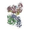
| ||||||||
|---|---|---|---|---|---|---|---|---|---|
| 1 | 
| ||||||||
| 2 | 
| ||||||||
| Unit cell |
|
-Components
-Protein , 2 types, 4 molecules ACBD
| #1: Protein | Mass: 64022.414 Da / Num. of mol.: 2 / Mutation: Q258C, C280S, D498N Source method: isolated from a genetically manipulated source Source: (gene. exp.) Human immunodeficiency virus type 1 BH10Gene: gag-pol / Production host:  References: UniProt: P03366, HIV-1 retropepsin, RNA-directed DNA polymerase, DNA-directed DNA polymerase, retroviral ribonuclease H, exoribonuclease H, Transferases; Transferring phosphorus- ...References: UniProt: P03366, HIV-1 retropepsin, RNA-directed DNA polymerase, DNA-directed DNA polymerase, retroviral ribonuclease H, exoribonuclease H, Transferases; Transferring phosphorus-containing groups; Nucleotidyltransferases, Hydrolases; Acting on ester bonds #2: Protein | Mass: 51928.629 Da / Num. of mol.: 2 / Mutation: C280S Source method: isolated from a genetically manipulated source Source: (gene. exp.) Human immunodeficiency virus type 1 BH10Gene: gag-pol / Production host: References: UniProt: P03366, HIV-1 retropepsin, RNA-directed DNA polymerase, DNA-directed DNA polymerase, retroviral ribonuclease H, exoribonuclease H, Transferases; Transferring phosphorus- ...References: UniProt: P03366, HIV-1 retropepsin, RNA-directed DNA polymerase, DNA-directed DNA polymerase, retroviral ribonuclease H, exoribonuclease H, Transferases; Transferring phosphorus-containing groups; Nucleotidyltransferases, Hydrolases; Acting on ester bonds |
|---|
-RNA chain , 2 types, 4 molecules TEPF
| #3: RNA chain | Mass: 7519.592 Da / Num. of mol.: 2 / Source method: obtained synthetically / Details: pbs SEQUENCE / Source: (synth.) Human immunodeficiency virus 1#4: RNA chain | Mass: 5331.189 Da / Num. of mol.: 2 / Source method: obtained synthetically / Details: tRNA(LYS3) / Source: (synth.) Homo sapiens (human) |
|---|
-Sugars , 1 types, 1 molecules
| #5: Polysaccharide | beta-D-fructofuranose-(2-1)-alpha-D-glucopyranose / sucrose  Source method: isolated from a genetically manipulated source Details: oligosaccharide with reducing-end-to-reducing-end glycosidic bond References: sucrose |
|---|
-Non-polymers , 2 types, 2 molecules 

| #6: Chemical | ChemComp-MG /  Mass: 24.305 Da / Num. of mol.: 1 Mass: 24.305 Da / Num. of mol.: 1Source method: isolated from a genetically manipulated source Formula: Mg |
|---|---|
| #7: Water | ChemComp-HOH / Mass: 18.015 Da / Num. of mol.: 1 / Source method: isolated from a natural source / Formula: H2O |
-Experimental details
-Experiment
| Experiment | Method: X-RAY DIFFRACTION / Number of used crystals: 1 |
|---|
- Sample preparation
| Crystal | Density Matthews: 3.39 Å3/Da / Density % sol: 63.72 % |
|---|---|
| Crystal grow | Temperature: 277 K / Method: vapor diffusion, sitting drop Details: PEG 8000, AMMONIUM SULFATE, MGCL2, GLYCEROL, SUCROSE PH range: 6.8 - 7.2 |
-Data collection
| Diffraction | Mean temperature: 100 K / Serial crystal experiment: N |
|---|---|
| Diffraction source | Source: SYNCHROTRON / Site: CHESS / Beamline: F1 / Wavelength: 0.9179 Å |
| Detector | Type: DECTRIS PILATUS3 S 6M / Detector: PIXEL / Date: Mar 28, 2014 |
| Radiation | Protocol: SINGLE WAVELENGTH / Monochromatic (M) / Laue (L): M / Scattering type: x-ray |
| Radiation wavelength | Wavelength: 0.9179 Å / Relative weight: 1 |
| Reflection | Resolution: 3.95→87.62 Å / Num. obs: 31549 / % possible obs: 99.9 % / Redundancy: 10.8 % / Biso Wilson estimate: 159.27 Å2 / Rmerge(I) obs: 0.236 / Net I/σ(I): 6.7 |
| Reflection shell | Resolution: 3.95→4.16 Å / Redundancy: 10.1 % / Rmerge(I) obs: 2.446 / Mean I/σ(I) obs: 0.9 / % possible all: 100 |
- Processing
| Software |
| ||||||||||||||||||||||||||||||||||||||||||||||||||||||||||||||||||||||||||||||||||||
|---|---|---|---|---|---|---|---|---|---|---|---|---|---|---|---|---|---|---|---|---|---|---|---|---|---|---|---|---|---|---|---|---|---|---|---|---|---|---|---|---|---|---|---|---|---|---|---|---|---|---|---|---|---|---|---|---|---|---|---|---|---|---|---|---|---|---|---|---|---|---|---|---|---|---|---|---|---|---|---|---|---|---|---|---|---|
| Refinement | Method to determine structure: MOLECULAR REPLACEMENT Starting model: 5TXL Resolution: 3.95→87.62 Å / SU ML: 1.02 / Cross valid method: THROUGHOUT / σ(F): 1.33 / Phase error: 45.86 / Stereochemistry target values: ML
| ||||||||||||||||||||||||||||||||||||||||||||||||||||||||||||||||||||||||||||||||||||
| Solvent computation | Shrinkage radii: 0.9 Å / VDW probe radii: 1.11 Å / Solvent model: FLAT BULK SOLVENT MODEL | ||||||||||||||||||||||||||||||||||||||||||||||||||||||||||||||||||||||||||||||||||||
| Refinement step | Cycle: LAST / Resolution: 3.95→87.62 Å
| ||||||||||||||||||||||||||||||||||||||||||||||||||||||||||||||||||||||||||||||||||||
| Refine LS restraints |
| ||||||||||||||||||||||||||||||||||||||||||||||||||||||||||||||||||||||||||||||||||||
| LS refinement shell |
|
