 Movie
Movie Controller

[English] 日本語
 Yorodumi
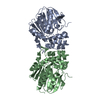
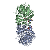
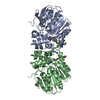
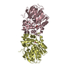
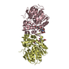
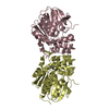
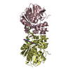
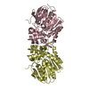
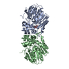
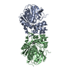
- PDB-5tne: Crystal structure of the E153Q mutant of the CFTR inhibitory fact... -
+ Open data

- Basic information
| Entry | Database: PDB / ID: 5tne | ||||||||||||||||||
|---|---|---|---|---|---|---|---|---|---|---|---|---|---|---|---|---|---|---|---|
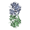
| Title | Crystal structure of the E153Q mutant of the CFTR inhibitory factor Cif containing the adducted cis-Stilbene Oxide hydrolysis intermediate | ||||||||||||||||||
 Components Components | CFTR inhibitory factor | ||||||||||||||||||
 Keywords Keywords | HYDROLASE / epoxide hydrolase / hydroxyalkyl-enzyme intermediate | ||||||||||||||||||
| Function / homology |  Function and homology information Function and homology information | ||||||||||||||||||
| Biological species |   Pseudomonas aeruginosa (bacteria) Pseudomonas aeruginosa (bacteria) | ||||||||||||||||||
| Method |  X-RAY DIFFRACTION / SYNCHROTRON / MOLECULAR REPLACEMENT / Resolution: 1.75 Å X-RAY DIFFRACTION / SYNCHROTRON / MOLECULAR REPLACEMENT / Resolution: 1.75 Å | ||||||||||||||||||
 Authors Authors | Hvorecny, K.L. / Madden, D.R. | ||||||||||||||||||
| Funding support |  United States, 5items United States, 5items
| ||||||||||||||||||
 Citation Citation | Journal: Structure / Year: 2017 Title: Active-Site Flexibility and Substrate Specificity in a Bacterial Virulence Factor: Crystallographic Snapshots of an Epoxide Hydrolase. Authors: Hvorecny, K.L. / Bahl, C.D. / Kitamura, S. / Lee, K.S.S. / Hammock, B.D. / Morisseau, C. / Madden, D.R. | ||||||||||||||||||
| History |
|
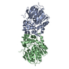
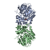
- Structure visualization
| Structure viewer | Molecule: MolmilJmol/JSmol |
|---|
- Downloads & links
-Download
| PDBx/mmCIF format | 5tne.cif.gz | 494.4 KB | Display | PDBx/mmCIF format |
|---|---|---|---|---|
| PDB format | pdb5tne.ent.gz | 406.3 KB | Display | PDB format |
| PDBx/mmJSON format | 5tne.json.gz | Tree view | PDBx/mmJSON format | |
| Others |  Other downloads Other downloads |
-Validation report
| Arichive directory | https://data.pdbj.org/pub/pdb/validation_reports/tn/5tneftp://data.pdbj.org/pub/pdb/validation_reports/tn/5tne | HTTPS FTP |
|---|
-Related structure data
| Related structure data | 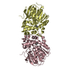 5tndC 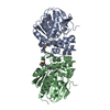 5tnfC 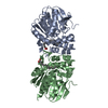 5tngC 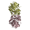 5tnhC 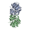 5tniC  5tnjC 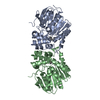 5tnkC 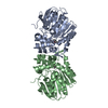 5tnlC  5tnmC 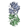 5tnnC  5tnpC  5tnqC  5tnrC  5tnsC  3kd2S S: Starting model for refinement C: citing same article ( |
|---|---|
| Similar structure data |


-Links
 PDBj
PDBj

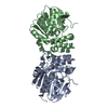
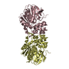
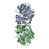
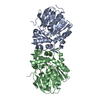
- Assembly
| Deposited unit | 
| |||||||||
|---|---|---|---|---|---|---|---|---|---|---|
| 1 | 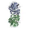
| |||||||||
| 2 | 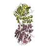
| |||||||||
| Unit cell |
| |||||||||
| Components on special symmetry positions |
|
-Components
| #1: Protein | Mass: 34163.715 Da / Num. of mol.: 4 / Mutation: E153Q Source method: isolated from a genetically manipulated source Source: (gene. exp.) Pseudomonas aeruginosa (strain UCBPP-PA14) (bacteria)Strain: UCBPP-PA14 / Gene: PA14_26090 / Production host: #2: Chemical |   Mass: 214.260 Da / Num. of mol.: 2 Mass: 214.260 Da / Num. of mol.: 2Source method: isolated from a genetically manipulated source Formula: C14H14O2 #3: Water | ChemComp-HOH / |  Mass: 18.015 Da / Num. of mol.: 1112 / Source method: isolated from a natural source / Formula: H2O Mass: 18.015 Da / Num. of mol.: 1112 / Source method: isolated from a natural source / Formula: H2OHas protein modification | Y | Nonpolymer details | 7F6 is the bound form ( covalently linked via OD1 atom of ASP 129 of CIF) of cis-Stilbene epoxide | |
|---|
-Experimental details
-Experiment
| Experiment | Method: X-RAY DIFFRACTION / Number of used crystals: 1 |
|---|
- Sample preparation
| Crystal | Density Matthews: 2.26 Å3/Da / Density % sol: 45.48 % |
|---|---|
| Crystal grow | Temperature: 291 K / Method: vapor diffusion, hanging drop / pH: 5 / Details: sodium acetate, pH5 calcium chloride PEG 8000 |
-Data collection
| Diffraction | Mean temperature: 100 K |
|---|---|
| Diffraction source | Source: SYNCHROTRON / Site: NSLS / Beamline: X6A / Wavelength: 0.9795 Å |
| Detector | Type: ADSC QUANTUM 270 / Detector: CCD / Date: Apr 20, 2016 |
| Radiation | Protocol: SINGLE WAVELENGTH / Monochromatic (M) / Laue (L): M / Scattering type: x-ray |
| Radiation wavelength | Wavelength: 0.9795 Å / Relative weight: 1 |
| Reflection | Resolution: 1.75→36.554 Å / Num. obs: 122215 / % possible obs: 99.8 % / Redundancy: 3.94 % / Rmerge(I) obs: 0.057 / Net I/σ(I): 19.62 |
| Reflection shell | Resolution: 1.75→1.8 Å / Redundancy: 3.91 % / Rmerge(I) obs: 0.357 / Mean I/σ(I) obs: 4.53 / % possible all: 99.8 |
- Processing
| Software |
| |||||||||||||||||||||||||||||||||||||||||||||||||||||||||||||||||||||||||||||||||||||||||||||||||||||||||||||||||||||||||||||||||||||||||||||||||||||||||||||||||||||||||||||||||||||||||||||||||||||||||||||||||||||||||
|---|---|---|---|---|---|---|---|---|---|---|---|---|---|---|---|---|---|---|---|---|---|---|---|---|---|---|---|---|---|---|---|---|---|---|---|---|---|---|---|---|---|---|---|---|---|---|---|---|---|---|---|---|---|---|---|---|---|---|---|---|---|---|---|---|---|---|---|---|---|---|---|---|---|---|---|---|---|---|---|---|---|---|---|---|---|---|---|---|---|---|---|---|---|---|---|---|---|---|---|---|---|---|---|---|---|---|---|---|---|---|---|---|---|---|---|---|---|---|---|---|---|---|---|---|---|---|---|---|---|---|---|---|---|---|---|---|---|---|---|---|---|---|---|---|---|---|---|---|---|---|---|---|---|---|---|---|---|---|---|---|---|---|---|---|---|---|---|---|---|---|---|---|---|---|---|---|---|---|---|---|---|---|---|---|---|---|---|---|---|---|---|---|---|---|---|---|---|---|---|---|---|---|---|---|---|---|---|---|---|---|---|---|---|---|---|---|---|---|
| Refinement | Method to determine structure: MOLECULAR REPLACEMENT Starting model: 3KD2 Resolution: 1.75→36.554 Å / SU ML: 0.17 / Cross valid method: FREE R-VALUE / σ(F): 1.99 / Phase error: 18
| |||||||||||||||||||||||||||||||||||||||||||||||||||||||||||||||||||||||||||||||||||||||||||||||||||||||||||||||||||||||||||||||||||||||||||||||||||||||||||||||||||||||||||||||||||||||||||||||||||||||||||||||||||||||||
| Solvent computation | Shrinkage radii: 0.9 Å / VDW probe radii: 1.11 Å | |||||||||||||||||||||||||||||||||||||||||||||||||||||||||||||||||||||||||||||||||||||||||||||||||||||||||||||||||||||||||||||||||||||||||||||||||||||||||||||||||||||||||||||||||||||||||||||||||||||||||||||||||||||||||
| Refinement step | Cycle: LAST / Resolution: 1.75→36.554 Å
| |||||||||||||||||||||||||||||||||||||||||||||||||||||||||||||||||||||||||||||||||||||||||||||||||||||||||||||||||||||||||||||||||||||||||||||||||||||||||||||||||||||||||||||||||||||||||||||||||||||||||||||||||||||||||
| Refine LS restraints |
| |||||||||||||||||||||||||||||||||||||||||||||||||||||||||||||||||||||||||||||||||||||||||||||||||||||||||||||||||||||||||||||||||||||||||||||||||||||||||||||||||||||||||||||||||||||||||||||||||||||||||||||||||||||||||
| LS refinement shell |
| |||||||||||||||||||||||||||||||||||||||||||||||||||||||||||||||||||||||||||||||||||||||||||||||||||||||||||||||||||||||||||||||||||||||||||||||||||||||||||||||||||||||||||||||||||||||||||||||||||||||||||||||||||||||||
| Refinement TLS params. | Method: refined / Refine-ID: X-RAY DIFFRACTION
| |||||||||||||||||||||||||||||||||||||||||||||||||||||||||||||||||||||||||||||||||||||||||||||||||||||||||||||||||||||||||||||||||||||||||||||||||||||||||||||||||||||||||||||||||||||||||||||||||||||||||||||||||||||||||
| Refinement TLS group |
|
