 Movie
Movie Controller

[English] 日本語
 Yorodumi
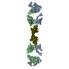
- PDB-5szo: Protocadherin Gamma B7 extracellular cadherin domains 1-4 P41212 ... -
+ Open data

- Basic information
| Entry | Database: PDB / ID: 5szo | ||||||||||||
|---|---|---|---|---|---|---|---|---|---|---|---|---|---|
| Title | Protocadherin Gamma B7 extracellular cadherin domains 1-4 P41212 crystal form | ||||||||||||
 Components Components | Protocadherin Gamma B7 | ||||||||||||
 Keywords Keywords | CELL ADHESION | ||||||||||||
| Function / homology |  Function and homology information Function and homology informationhomophilic cell-cell adhesion / cell adhesion molecule binding / cell adhesion / calcium ion binding / membrane / plasma membrane Similarity search - Function | ||||||||||||
| Biological species |  | ||||||||||||
| Method |  X-RAY DIFFRACTION / SYNCHROTRON / MOLECULAR REPLACEMENT / Resolution: 3.612 Å X-RAY DIFFRACTION / SYNCHROTRON / MOLECULAR REPLACEMENT / Resolution: 3.612 Å | ||||||||||||
 Authors Authors | Goodman, K.M. / Mannepalli, S. / Bahna, F. / Honig, B. / Shapiro, L. | ||||||||||||
| Funding support |  United States, 3items United States, 3items
| ||||||||||||
 Citation Citation | Journal: Elife / Year: 2016 Title: gamma-Protocadherin structural diversity and functional implications. Authors: Goodman, K.M. / Rubinstein, R. / Thu, C.A. / Mannepalli, S. / Bahna, F. / Ahlsen, G. / Rittenhouse, C. / Maniatis, T. / Honig, B. / Shapiro, L. | ||||||||||||
| History |
|
- Structure visualization
| Structure viewer | Molecule: MolmilJmol/JSmol |
|---|
- Downloads & links
-Download
| PDBx/mmCIF format | 5szo.cif.gz | 178.7 KB | Display | PDBx/mmCIF format |
|---|---|---|---|---|
| PDB format | pdb5szo.ent.gz | 137.3 KB | Display | PDB format |
| PDBx/mmJSON format | 5szo.json.gz | Tree view | PDBx/mmJSON format | |
| Others |  Other downloads Other downloads |
-Validation report
| Arichive directory | https://data.pdbj.org/pub/pdb/validation_reports/sz/5szoftp://data.pdbj.org/pub/pdb/validation_reports/sz/5szo | HTTPS FTP |
|---|
-Related structure data
| Related structure data | 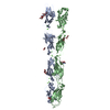 5szlC 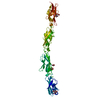 5szmC 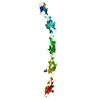 5sznSC  5szpC 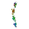 5szqC 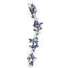 5szrC 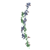 5t9tC  4zi8S  4zi9S  4zplS  4zpmS 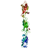 4zpoS  4zpsS  5dzvS  5dzxS  5zsmS C: citing same article ( S: Starting model for refinement |
|---|---|
| Similar structure data |
-Links
 PDBj
PDBj


- Assembly
| Deposited unit | 
| ||||||||
|---|---|---|---|---|---|---|---|---|---|
| 1 |
| ||||||||
| Unit cell |
| ||||||||
| Details | The oligomeric state was determined by sedimentation equilibrium analytical ultracentrifugation |
-Components
| #1: Protein | Mass: 47295.547 Da / Num. of mol.: 2 Source method: isolated from a genetically manipulated source Source: (gene. exp.)  Homo sapiens (human) / References: UniProt: Q91XX3 Homo sapiens (human) / References: UniProt: Q91XX3#2: Polysaccharide | alpha-L-fucopyranose-(1-6)-2-acetamido-2-deoxy-beta-D-glucopyranose | Source method: isolated from a genetically manipulated source #3: Sugar | ChemComp-MAN /   Type: D-saccharide, alpha linking / Mass: 180.156 Da / Num. of mol.: 8 Type: D-saccharide, alpha linking / Mass: 180.156 Da / Num. of mol.: 8Source method: isolated from a genetically manipulated source Formula: C6H12O6 #4: Chemical | ChemComp-CA /   Mass: 40.078 Da / Num. of mol.: 18 / Source method: obtained synthetically / Formula: Ca Mass: 40.078 Da / Num. of mol.: 18 / Source method: obtained synthetically / Formula: CaHas protein modification | Y | |
|---|
-Experimental details
-Experiment
| Experiment | Method: X-RAY DIFFRACTION / Number of used crystals: 1 |
|---|
- Sample preparation
| Crystal | Density Matthews: 3.9 Å3/Da / Density % sol: 68.43 % |
|---|---|
| Crystal grow | Temperature: 295 K / Method: batch mode / pH: 8.5 Details: 0.1 M Tris-Cl pH 8.5, 0.2 M trimethylamine N-oxide, 3% dextran sulfate sodium salt 5000, 17% (w/v) PEG2000MME |
-Data collection
| Diffraction | Mean temperature: 100 K |
|---|---|
| Diffraction source | Source: SYNCHROTRON / Site: APS / Beamline: 24-ID-C / Wavelength: 0.9793 Å |
| Detector | Type: DECTRIS PILATUS 6M-F / Detector: PIXEL / Date: Oct 25, 2015 |
| Radiation | Protocol: SINGLE WAVELENGTH / Monochromatic (M) / Laue (L): M / Scattering type: x-ray |
| Radiation wavelength | Wavelength: 0.9793 Å / Relative weight: 1 |
| Reflection | Resolution: 3.59→104.13 Å / Num. obs: 18347 / % possible obs: 99.6 % / Redundancy: 12.5 % / Biso Wilson estimate: 109.88 Å2 / CC1/2: 1 / Rmerge(I) obs: 0.183 / Net I/σ(I): 9 |
| Reflection shell | Resolution: 3.59→3.93 Å / Redundancy: 12.3 % / Rmerge(I) obs: 3.722 / Mean I/σ(I) obs: 1 / CC1/2: 0.741 / % possible all: 98.7 |
- Processing
| Software |
| |||||||||||||||||||||||||||||||||||
|---|---|---|---|---|---|---|---|---|---|---|---|---|---|---|---|---|---|---|---|---|---|---|---|---|---|---|---|---|---|---|---|---|---|---|---|---|
| Refinement | Method to determine structure: MOLECULAR REPLACEMENT Starting model: 4ZPL, 4ZPM, 4ZPO, 4ZPS, 5DZV, 5DZX, 4ZI9, 4ZI8, 5ZSM, and 5SZN Resolution: 3.612→19.93 Å / SU ML: 0.5 / Cross valid method: FREE R-VALUE / σ(F): 1.36 / Phase error: 44.17 Details: Ellipsoidal resolution limits were applied to the data due to severe anisotropy. The limits are 4.5/4.5/3.6 angstrom along the a*/b*/c* axes respectively. Completeness with respect to the ...Details: Ellipsoidal resolution limits were applied to the data due to severe anisotropy. The limits are 4.5/4.5/3.6 angstrom along the a*/b*/c* axes respectively. Completeness with respect to the diffracting ellipsoid = 90%.
| |||||||||||||||||||||||||||||||||||
| Solvent computation | Shrinkage radii: 0.9 Å / VDW probe radii: 1.11 Å | |||||||||||||||||||||||||||||||||||
| Refinement step | Cycle: LAST / Resolution: 3.612→19.93 Å
| |||||||||||||||||||||||||||||||||||
| Refine LS restraints |
| |||||||||||||||||||||||||||||||||||
| LS refinement shell |
|
