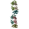
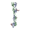
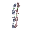
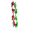
[English] 日本語
Yorodumi
- PDB-6bx7: Crystal Structure of Human Protocadherin-1 EC1-4 -

+
Open data


ID or keywords:

Loading...

-
Basic information

Entry
Database: PDB / ID: 6bx7
TitleCrystal Structure of Human Protocadherin-1 EC1-4
ComponentsProtocadherin-1
KeywordsCELL ADHESION / cadherins / adhesion / calcium-binding protein / asthma
Function / homology
Function and homology information


homophilic cell adhesion via plasma membrane adhesion molecules / cell-cell junction / cell-cell signaling / cell junction / nervous system development / cell adhesion / intracellular membrane-bounded organelle / calcium ion binding / nucleolus / nucleoplasm / plasma membrane
Similarity search - Function
Protocadherin / Protocadherin / Cadherin, N-terminal / Cadherin-like / Cadherins / Cadherin conserved site / Cadherin domain signature. / Cadherin repeats. / Cadherin domain / Cadherins domain profile. ...Protocadherin / Protocadherin / Cadherin, N-terminal / Cadherin-like / Cadherins / Cadherin conserved site / Cadherin domain signature. / Cadherin repeats. / Cadherin domain / Cadherins domain profile. / Cadherin-like / Cadherin-like superfamily / Immunoglobulin-like / Sandwich / Mainly Beta
Similarity search - Domain/homology
Biological speciesHomo sapiens (human)
MethodX-RAY DIFFRACTION / SYNCHROTRON / MOLECULAR REPLACEMENT / Resolution: 2.85 Å
AuthorsModak, D. / Sotomayor, M.
Funding support United States, 1items
OrganizationGrant numberCountry
Other privateAlfred P. Sloan FR-2015-65794 United States
CitationJournal: Commun Biol / Year: 2019
Title: Identification of an adhesive interface for the non-clustered delta 1 protocadherin-1 involved in respiratory diseases.
Authors: Modak, D. / Sotomayor, M.
History
DepositionDec 17, 2017Deposition site: RCSB / Processing site: RCSB
Revision 1.0Dec 19, 2018Provider: repository / Type: Initial release
Revision 1.1Oct 9, 2019Group: Data collection / Database references / Category: citation
Item: _citation.country / _citation.journal_abbrev ..._citation.country / _citation.journal_abbrev / _citation.journal_id_CSD / _citation.journal_id_ISSN / _citation.pdbx_database_id_DOI / _citation.title / _citation.year
Revision 1.2Oct 16, 2019Group: Data collection / Database references / Category: citation / citation_author
Item: _citation.journal_volume / _citation.page_first ..._citation.journal_volume / _citation.page_first / _citation.page_last / _citation.pdbx_database_id_PubMed / _citation.title / _citation_author.identifier_ORCID
Revision 1.3Oct 4, 2023Group: Data collection / Database references ...Data collection / Database references / Derived calculations / Refinement description
Category: chem_comp_atom / chem_comp_bond ...chem_comp_atom / chem_comp_bond / database_2 / pdbx_initial_refinement_model / pdbx_struct_conn_angle / struct_conn
Item: _database_2.pdbx_DOI / _database_2.pdbx_database_accession ..._database_2.pdbx_DOI / _database_2.pdbx_database_accession / _pdbx_struct_conn_angle.ptnr1_auth_comp_id / _pdbx_struct_conn_angle.ptnr1_auth_seq_id / _pdbx_struct_conn_angle.ptnr1_label_asym_id / _pdbx_struct_conn_angle.ptnr1_label_atom_id / _pdbx_struct_conn_angle.ptnr1_label_comp_id / _pdbx_struct_conn_angle.ptnr1_label_seq_id / _pdbx_struct_conn_angle.ptnr2_auth_seq_id / _pdbx_struct_conn_angle.ptnr2_label_asym_id / _pdbx_struct_conn_angle.ptnr3_auth_comp_id / _pdbx_struct_conn_angle.ptnr3_auth_seq_id / _pdbx_struct_conn_angle.ptnr3_label_asym_id / _pdbx_struct_conn_angle.ptnr3_label_atom_id / _pdbx_struct_conn_angle.ptnr3_label_comp_id / _pdbx_struct_conn_angle.ptnr3_label_seq_id / _pdbx_struct_conn_angle.value / _struct_conn.pdbx_dist_value / _struct_conn.ptnr1_auth_comp_id / _struct_conn.ptnr1_auth_seq_id / _struct_conn.ptnr1_label_asym_id / _struct_conn.ptnr1_label_atom_id / _struct_conn.ptnr1_label_comp_id / _struct_conn.ptnr1_label_seq_id / _struct_conn.ptnr2_auth_comp_id / _struct_conn.ptnr2_auth_seq_id / _struct_conn.ptnr2_label_asym_id / _struct_conn.ptnr2_label_atom_id / _struct_conn.ptnr2_label_comp_id / _struct_conn.ptnr2_symmetry

-
Structure visualization

Structure viewerMolecule:
MolmilJmol/JSmol

Downloads & links

-
Assembly

Deposited unit
A: Protocadherin-1
hetero molecules


Theoretical massNumber of molelcules
Total (without water)50,48113
Polymers49,8021
Non-polymers67912
Water45025
1
A: Protocadherin-1
hetero molecules

A: Protocadherin-1
hetero molecules


Theoretical massNumber of molelcules
Total (without water)100,96226
Polymers99,6042
Non-polymers1,35824
Water362
TypeNameSymmetry operationNumber
identity operation1_555x,y,z1
crystal symmetry operation8_555x-y,-y,-z1
Buried area4850 Å2
ΔGint-118 kcal/mol
Surface area46740 Å2
MethodPISA
Unit cell
Length a, b, c (Å)146.981, 146.981, 147.686
Angle α, β, γ (deg.)90.00, 90.00, 120.00
Int Tables number178
Space group name H-MP6122

-
Components

#1: Protein Protocadherin-1 / / Cadherin-like protein 1 / Protocadherin-42 / PC42


Mass: 49801.922 Da / Num. of mol.: 1 / Fragment: Cadherin domains 1-4, residues 58-503
Source method: isolated from a genetically manipulated source
Source: (gene. exp.) Homo sapiens (human) / Gene: PCDH1 / Plasmid: PET21A / Production host: Escherichia coli BL21(DE3) (bacteria) / References: UniProt: Q08174
#2: Chemical ChemComp-EPE / 4-(2-HYDROXYETHYL)-1-PIPERAZINE ETHANESULFONIC ACID / HEPES / HEPES


Mass: 238.305 Da / Num. of mol.: 1 / Source method: obtained synthetically / Formula: C8H18N2O4S / Comment: pH buffer*YM
#3: Chemical
ChemComp-CA / CALCIUM ION


Mass: 40.078 Da / Num. of mol.: 11 / Source method: obtained synthetically / Formula: Ca
#4: Water ChemComp-HOH / water / Water


Mass: 18.015 Da / Num. of mol.: 25 / Source method: isolated from a natural source / Formula: H2O

-
Experimental details

-
Experiment

ExperimentMethod: X-RAY DIFFRACTION / Number of used crystals: 1

-
Sample preparation

CrystalDensity Matthews: 4.63 Å3/Da / Density % sol: 73.42 %
Crystal growTemperature: 277 K / Method: vapor diffusion, sitting drop / pH: 7.5
Details: 0.2 M Calcium Chloride, 0.1 M HEPES Sodium Salt pH 7.5, 15% PEG 400 + 15% Glycerol

-
Data collection

DiffractionMean temperature: 100 K
Diffraction sourceSource: SYNCHROTRON / Site: APS / Beamline: 24-ID-E / Wavelength: 0.97921 Å
DetectorType: ADSC QUANTUM 315 / Detector: CCD / Date: Dec 18, 2016
RadiationProtocol: SINGLE WAVELENGTH / Monochromatic (M) / Laue (L): M / Scattering type: x-ray
Radiation wavelengthWavelength: 0.97921 Å / Relative weight: 1
ReflectionResolution: 2.85→50 Å / Num. obs: 22567 / % possible obs: 100 % / Redundancy: 10.7 % / CC1/2: 0.998 / Rmerge(I) obs: 0.156 / Rpim(I) all: 0.05 / Rrim(I) all: 0.164 / Net I/σ(I): 18.3
Reflection shellResolution: 2.85→2.9 Å / Redundancy: 10.6 % / Rmerge(I) obs: 1.627 / Mean I/σ(I) obs: 1.92 / Num. unique obs: 1103 / CC1/2: 0.716 / Rpim(I) all: 0.521 / Rrim(I) all: 1.71 / % possible all: 100

-
Processing

Software
NameVersionClassification
REFMAC5.8.0230refinement
HKL-2000data reduction
HKL-2000data scaling
PHASERphasing
RefinementMethod to determine structure: MOLECULAR REPLACEMENT
Starting model: 5DZV
Resolution: 2.85→48.26 Å / Cor.coef. Fo:Fc: 0.929 / Cor.coef. Fo:Fc free: 0.926 / SU B: 26.155 / SU ML: 0.224 / Cross valid method: THROUGHOUT / ESU R: 0.413 / ESU R Free: 0.279 / Details: HYDROGENS HAVE BEEN ADDED IN THE RIDING POSITIONS
RfactorNum. reflection% reflectionSelection details
Rfree0.24336 1120 5 %RANDOM
Rwork0.22097 ---
obs0.22213 21392 100 %-
Solvent computationIon probe radii: 0.8 Å / Shrinkage radii: 0.8 Å / VDW probe radii: 1.2 Å
Displacement parametersBiso mean: 75.792 Å2
Baniso -1Baniso -2Baniso -3
1-1.04 Å20.52 Å20 Å2
2--1.04 Å2-0 Å2
3----3.36 Å2
Refinement stepCycle: 1 / Resolution: 2.85→48.26 Å
ProteinNucleic acidLigandSolventTotal
Num. atoms3288 0 26 25 3339
Refine LS restraints
Refine-IDTypeDev idealDev ideal targetNumber
X-RAY DIFFRACTIONr_bond_refined_d0.010.0143354
X-RAY DIFFRACTIONr_bond_other_d0.0010.0172996
X-RAY DIFFRACTIONr_angle_refined_deg1.4841.6664569
X-RAY DIFFRACTIONr_angle_other_deg0.8861.6417065
X-RAY DIFFRACTIONr_dihedral_angle_1_deg8.7485426
X-RAY DIFFRACTIONr_dihedral_angle_2_deg38.97624.098183
X-RAY DIFFRACTIONr_dihedral_angle_3_deg17.94415557
X-RAY DIFFRACTIONr_dihedral_angle_4_deg23.4431521
X-RAY DIFFRACTIONr_chiral_restr0.0660.2455
X-RAY DIFFRACTIONr_gen_planes_refined0.0050.023799
X-RAY DIFFRACTIONr_gen_planes_other0.0010.02521
X-RAY DIFFRACTIONr_nbd_refined
X-RAY DIFFRACTIONr_nbd_other
X-RAY DIFFRACTIONr_nbtor_refined
X-RAY DIFFRACTIONr_nbtor_other
X-RAY DIFFRACTIONr_xyhbond_nbd_refined
X-RAY DIFFRACTIONr_xyhbond_nbd_other
X-RAY DIFFRACTIONr_metal_ion_refined
X-RAY DIFFRACTIONr_metal_ion_other
X-RAY DIFFRACTIONr_symmetry_vdw_refined
X-RAY DIFFRACTIONr_symmetry_vdw_other
X-RAY DIFFRACTIONr_symmetry_hbond_refined
X-RAY DIFFRACTIONr_symmetry_hbond_other
X-RAY DIFFRACTIONr_symmetry_metal_ion_refined
X-RAY DIFFRACTIONr_symmetry_metal_ion_other
X-RAY DIFFRACTIONr_mcbond_it2.0964.4741713
X-RAY DIFFRACTIONr_mcbond_other2.0684.471712
X-RAY DIFFRACTIONr_mcangle_it3.6086.72136
X-RAY DIFFRACTIONr_mcangle_other3.6116.7062137
X-RAY DIFFRACTIONr_scbond_it1.9144.7491640
X-RAY DIFFRACTIONr_scbond_other1.9134.751640
X-RAY DIFFRACTIONr_scangle_it
X-RAY DIFFRACTIONr_scangle_other3.2577.0342434
X-RAY DIFFRACTIONr_long_range_B_refined8.7384.15613155
X-RAY DIFFRACTIONr_long_range_B_other8.72984.15213146
X-RAY DIFFRACTIONr_rigid_bond_restr
X-RAY DIFFRACTIONr_sphericity_free
X-RAY DIFFRACTIONr_sphericity_bonded
LS refinement shellResolution: 2.851→2.925 Å / Total num. of bins used: 20
RfactorNum. reflection% reflection
Rfree0.372 91 -
Rwork0.338 1517 -
obs--99.01 %
Refinement TLS params.

Method: refined / Refine-ID: X-RAY DIFFRACTION

IDL112)L122)L132)L222)L232)L332)S11 (Å °)S12 (Å °)S13 (Å °)S21 (Å °)S22 (Å °)S23 (Å °)S31 (Å °)S32 (Å °)S33 (Å °)T112)T122)T132)T222)T232)T332)Origin x (Å)Origin y (Å)Origin z (Å)
12.4115-1.1832-2.76724.11574.68556.60410.3065-0.3730.56790.10340.0894-0.0897-0.52570.0926-0.39580.69680.1459-0.09990.6092-0.06750.475557.682221.597760.3586
20.49970.35261.10570.41341.65828.21350.0233-0.02540.01250.03370.0215-0.09680.03620.1392-0.04480.17140.0207-0.0740.011-0.02060.287865.5707-0.406722.8339
32.4852.56153.08813.08324.10666.2596-0.06480.1388-0.0107-0.2620.07040.0197-0.3143-0.0704-0.00560.170.00110.01960.03720.01470.133448.3599-18.5947-19.7646
40.4853-0.1094-0.66740.82230.85737.55910.07420.441-0.0077-0.59860.020.0776-0.14560.0721-0.09420.5939-0.0613-0.04430.5364-0.05590.353238.6049-37.4569-62.2438
Refinement TLS group
IDRefine-IDRefine TLS-IDAuth asym-IDAuth seq-ID
1X-RAY DIFFRACTION1A3 - 106
2X-RAY DIFFRACTION1A502 - 503
3X-RAY DIFFRACTION2A107 - 218
4X-RAY DIFFRACTION2A504 - 506
5X-RAY DIFFRACTION3A219 - 325
6X-RAY DIFFRACTION3A507 - 509
7X-RAY DIFFRACTION4A326 - 439
8X-RAY DIFFRACTION4A510 - 511

+
About Yorodumi

-
News

-
Feb 9, 2022. New format data for meta-information of EMDB entries

New format data for meta-information of EMDB entries

  • Version 3 of the EMDB header file is now the official format.
  • The previous official version 1.9 will be removed from the archive.

Related info.:EMDB header

External links:wwPDB to switch to version 3 of the EMDB data model

-
Aug 12, 2020. Covid-19 info

Covid-19 info

URL: https://pdbj.org/emnavi/covid19.php

New page: Covid-19 featured information page in EM Navigator.

Related info.:Covid-19 info / Mar 5, 2020. Novel coronavirus structure data

+
Mar 5, 2020. Novel coronavirus structure data

Novel coronavirus structure data

Related info.:Yorodumi Speices / Aug 12, 2020. Covid-19 info

External links:COVID-19 featured content - PDBj / Molecule of the Month (242):Coronavirus Proteases

+
Jan 31, 2019. EMDB accession codes are about to change! (news from PDBe EMDB page)

EMDB accession codes are about to change! (news from PDBe EMDB page)

  • The allocation of 4 digits for EMDB accession codes will soon come to an end. Whilst these codes will remain in use, new EMDB accession codes will include an additional digit and will expand incrementally as the available range of codes is exhausted. The current 4-digit format prefixed with “EMD-” (i.e. EMD-XXXX) will advance to a 5-digit format (i.e. EMD-XXXXX), and so on. It is currently estimated that the 4-digit codes will be depleted around Spring 2019, at which point the 5-digit format will come into force.
  • The EM Navigator/Yorodumi systems omit the EMD- prefix.

Related info.:Q: What is EMD? / ID/Accession-code notation in Yorodumi/EM Navigator

External links:EMDB Accession Codes are Changing Soon! / Contact to PDBj

+
Jul 12, 2017. Major update of PDB

Major update of PDB

  • wwPDB released updated PDB data conforming to the new PDBx/mmCIF dictionary.
  • This is a major update changing the version number from 4 to 5, and with Remediation, in which all the entries are updated.
  • In this update, many items about electron microscopy experimental information are reorganized (e.g. em_software).
  • Now, EM Navigator and Yorodumi are based on the updated data.

External links:wwPDB Remediation / Enriched Model Files Conforming to OneDep Data Standards Now Available in the PDB FTP Archive

-
Yorodumi

Thousand views of thousand structures

  • Yorodumi is a browser for structure data from EMDB, PDB, SASBDB, etc.
  • This page is also the successor to EM Navigator detail page, and also detail information page/front-end page for Omokage search.
  • The word "yorodu" (or yorozu) is an old Japanese word meaning "ten thousand". "mi" (miru) is to see.

Related info.:EMDB / PDB / SASBDB / Comparison of 3 databanks / Yorodumi Search / Aug 31, 2016. New EM Navigator & Yorodumi / Yorodumi Papers / Jmol/JSmol / Function and homology information / Changes in new EM Navigator and Yorodumi

Read more