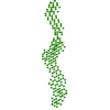
 Movie
Movie Controller


+ Open data

- Basic information
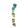
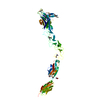
| Entry | Database: PDB / ID: 5szq | |||||||||
|---|---|---|---|---|---|---|---|---|---|---|
| Title | Protocadherin Gamma A4 extracellular cadherin domains 3-6 | |||||||||
 Components Components | Protocadherin gamma-A4 | |||||||||
 Keywords Keywords | CELL ADHESION | |||||||||
| Function / homology |  Function and homology information Function and homology informationhomophilic cell-cell adhesion / cell adhesion / calcium ion binding / membrane / plasma membrane Similarity search - Function | |||||||||
| Biological species |  | |||||||||
| Method |  X-RAY DIFFRACTION / SYNCHROTRON / MOLECULAR REPLACEMENT / Resolution: 2.608 Å X-RAY DIFFRACTION / SYNCHROTRON / MOLECULAR REPLACEMENT / Resolution: 2.608 Å | |||||||||
 Authors Authors | Goodman, K.M. / Mannepalli, S. / Bahna, F. / Honig, B. / Shapiro, L. | |||||||||
 Citation Citation | Journal: Elife / Year: 2016 Title: gamma-Protocadherin structural diversity and functional implications. Authors: Goodman, K.M. / Rubinstein, R. / Thu, C.A. / Mannepalli, S. / Bahna, F. / Ahlsen, G. / Rittenhouse, C. / Maniatis, T. / Honig, B. / Shapiro, L. | |||||||||
| History |
|
- Structure visualization
| Structure viewer | Molecule: MolmilJmol/JSmol |
|---|
- Downloads & links
-Download
| PDBx/mmCIF format | 5szq.cif.gz | 184.6 KB | Display | PDBx/mmCIF format |
|---|---|---|---|---|
| PDB format | pdb5szq.ent.gz | 146.9 KB | Display | PDB format |
| PDBx/mmJSON format | 5szq.json.gz | Tree view | PDBx/mmJSON format | |
| Others |  Other downloads Other downloads |
-Validation report
| Arichive directory | https://data.pdbj.org/pub/pdb/validation_reports/sz/5szqftp://data.pdbj.org/pub/pdb/validation_reports/sz/5szq | HTTPS FTP |
|---|
-Related structure data
| Related structure data |  5szlC  5szmSC  5sznSC  5szoC  5szpC  5szrSC  5t9tC S: Starting model for refinement C: citing same article ( |
|---|---|
| Similar structure data |

-Links
 PDBj
PDBj


- Assembly
| Deposited unit | 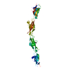
| ||||||||
|---|---|---|---|---|---|---|---|---|---|
| 1 |
| ||||||||
| Unit cell |
|
-Components
| #1: Protein | Mass: 48060.758 Da / Num. of mol.: 1 Source method: isolated from a genetically manipulated source Details: PcdhgA4 extracellular domains 3-6 with C-terminal octahistidine tag Source: (gene. exp.)  Homo sapiens (human) / References: UniProt: Q91XY4 Homo sapiens (human) / References: UniProt: Q91XY4 | ||||||
|---|---|---|---|---|---|---|---|
| #2: Polysaccharide | 2-acetamido-2-deoxy-beta-D-glucopyranose-(1-4)-2-acetamido-2-deoxy-beta-D-glucopyranose Source method: isolated from a genetically manipulated source | ||||||
| #3: Sugar | ChemComp-MAN /   Type: D-saccharide, alpha linking / Mass: 180.156 Da / Num. of mol.: 6 Type: D-saccharide, alpha linking / Mass: 180.156 Da / Num. of mol.: 6Source method: isolated from a genetically manipulated source Formula: C6H12O6 #4: Sugar | ChemComp-NAG / |   Type: D-saccharide, beta linking / Mass: 221.208 Da / Num. of mol.: 1 Type: D-saccharide, beta linking / Mass: 221.208 Da / Num. of mol.: 1Source method: isolated from a genetically manipulated source Formula: C8H15NO6 #5: Chemical | ChemComp-CA /   Mass: 40.078 Da / Num. of mol.: 9 / Source method: obtained synthetically / Formula: Ca Mass: 40.078 Da / Num. of mol.: 9 / Source method: obtained synthetically / Formula: CaHas protein modification | Y | |
-Experimental details
-Experiment
| Experiment | Method: X-RAY DIFFRACTION / Number of used crystals: 1 |
|---|
- Sample preparation
| Crystal | Density Matthews: 3.63 Å3/Da / Density % sol: 66.16 % |
|---|---|
| Crystal grow | Temperature: 295 K / Method: batch mode / pH: 8.5 Details: 0.1 M ammonium sulfate, 9% (w/v) PEG20000, 18% PEG550MME, 0.1 M Morpheus Buffer System 3 (Tris/Bicine; Molecular Dimensions) pH 8.5 |
-Data collection
| Diffraction | Mean temperature: 100 K |
|---|---|
| Diffraction source | Source: SYNCHROTRON / Site: APS  / Beamline: 24-ID-C / Wavelength: 0.97919 Å / Beamline: 24-ID-C / Wavelength: 0.97919 Å |
| Detector | Type: DECTRIS PILATUS 6M-F / Detector: PIXEL / Date: Jun 29, 2016 |
| Radiation | Protocol: SINGLE WAVELENGTH / Monochromatic (M) / Laue (L): M / Scattering type: x-ray |
| Radiation wavelength | Wavelength: 0.97919 Å / Relative weight: 1 |
| Reflection | Resolution: 2.56→172.16 Å / Num. obs: 23763 / % possible obs: 99.3 % / Redundancy: 4.1 % / Biso Wilson estimate: 48.68 Å2 / CC1/2: 0.998 / Rmerge(I) obs: 0.112 / Net I/σ(I): 6.6 |
| Reflection shell | Resolution: 2.56→2.67 Å / Redundancy: 3.3 % / Rmerge(I) obs: 3.118 / CC1/2: 0.434 / % possible all: 97.5 |
- Processing
| Software |
| |||||||||||||||||||||||||||||||||||||||||||||||||||||||||||||||||||||||||||||||||||||||||||||||||||||||||||||||||||||||||||||
|---|---|---|---|---|---|---|---|---|---|---|---|---|---|---|---|---|---|---|---|---|---|---|---|---|---|---|---|---|---|---|---|---|---|---|---|---|---|---|---|---|---|---|---|---|---|---|---|---|---|---|---|---|---|---|---|---|---|---|---|---|---|---|---|---|---|---|---|---|---|---|---|---|---|---|---|---|---|---|---|---|---|---|---|---|---|---|---|---|---|---|---|---|---|---|---|---|---|---|---|---|---|---|---|---|---|---|---|---|---|---|---|---|---|---|---|---|---|---|---|---|---|---|---|---|---|---|
| Refinement | Method to determine structure: MOLECULAR REPLACEMENT Starting model: EC3-4 of 5SZM, EC5 of 5SZN and EC6 of 5SZR Resolution: 2.608→19.957 Å / SU ML: 0.31 / Cross valid method: FREE R-VALUE / σ(F): 1.36 / Phase error: 43.82 / Stereochemistry target values: ML
| |||||||||||||||||||||||||||||||||||||||||||||||||||||||||||||||||||||||||||||||||||||||||||||||||||||||||||||||||||||||||||||
| Solvent computation | Shrinkage radii: 0.9 Å / VDW probe radii: 1.11 Å / Solvent model: FLAT BULK SOLVENT MODEL | |||||||||||||||||||||||||||||||||||||||||||||||||||||||||||||||||||||||||||||||||||||||||||||||||||||||||||||||||||||||||||||
| Refinement step | Cycle: LAST / Resolution: 2.608→19.957 Å
| |||||||||||||||||||||||||||||||||||||||||||||||||||||||||||||||||||||||||||||||||||||||||||||||||||||||||||||||||||||||||||||
| Refine LS restraints |
| |||||||||||||||||||||||||||||||||||||||||||||||||||||||||||||||||||||||||||||||||||||||||||||||||||||||||||||||||||||||||||||
| LS refinement shell |
| |||||||||||||||||||||||||||||||||||||||||||||||||||||||||||||||||||||||||||||||||||||||||||||||||||||||||||||||||||||||||||||
| Refinement TLS params. | Method: refined / Refine-ID: X-RAY DIFFRACTION
| |||||||||||||||||||||||||||||||||||||||||||||||||||||||||||||||||||||||||||||||||||||||||||||||||||||||||||||||||||||||||||||
| Refinement TLS group |
|
