 Movie
Movie Controller

[English] 日本語
 Yorodumi
- PDB-5lf5: Myelin-associated glycoprotein (MAG) deglycosylated full extracel... -
+ Open data

- Basic information
| Entry | Database: PDB / ID: 5lf5 | |||||||||
|---|---|---|---|---|---|---|---|---|---|---|
| Title | Myelin-associated glycoprotein (MAG) deglycosylated full extracellular domain with co-purified ligand | |||||||||
 Components Components | Myelin-associated glycoprotein | |||||||||
 Keywords Keywords | CELL ADHESION / Myelin / Signaling | |||||||||
| Function / homology |  Function and homology information Function and homology informationmesaxon / Axonal growth inhibition (RHOA activation) / compact myelin / Basigin interactions / ganglioside GT1b binding / myelin sheath adaxonal region / sialic acid binding / central nervous system myelin formation / : / negative regulation of axon extension ...mesaxon / Axonal growth inhibition (RHOA activation) / compact myelin / Basigin interactions / ganglioside GT1b binding / myelin sheath adaxonal region / sialic acid binding / central nervous system myelin formation / : / negative regulation of axon extension / central nervous system myelination / positive regulation of astrocyte differentiation / paranode region of axon / positive regulation of myelination / Schmidt-Lanterman incisure / axon regeneration / negative regulation of neuron differentiation / transmission of nerve impulse / myelination / cellular response to mechanical stimulus / myelin sheath / negative regulation of neuron projection development / carbohydrate binding / negative regulation of neuron apoptotic process / cell adhesion / membrane raft / signaling receptor binding / protein kinase binding / protein homodimerization activity / plasma membrane Similarity search - Function | |||||||||
| Biological species |  | |||||||||
| Method |  X-RAY DIFFRACTION / SYNCHROTRON / MOLECULAR REPLACEMENT / Resolution: 3.8 Å X-RAY DIFFRACTION / SYNCHROTRON / MOLECULAR REPLACEMENT / Resolution: 3.8 Å | |||||||||
 Authors Authors | Pronker, M.F. / Janssen, B.J.C. | |||||||||
| Funding support |  Netherlands, 1items Netherlands, 1items
| |||||||||
 Citation Citation | Journal: Nat Commun / Year: 2016 Title: Structural basis of myelin-associated glycoprotein adhesion and signalling. Authors: Matti F Pronker / Suzanne Lemstra / Joost Snijder / Albert J R Heck / Dominique M E Thies-Weesie / R Jeroen Pasterkamp / Bert J C Janssen / Abstract: Myelin-associated glycoprotein (MAG) is a myelin-expressed cell-adhesion and bi-directional signalling molecule. MAG maintains the myelin-axon spacing by interacting with specific neuronal ...Myelin-associated glycoprotein (MAG) is a myelin-expressed cell-adhesion and bi-directional signalling molecule. MAG maintains the myelin-axon spacing by interacting with specific neuronal glycolipids (gangliosides), inhibits axon regeneration and controls myelin formation. The mechanisms underlying MAG adhesion and signalling are unresolved. We present crystal structures of the MAG full ectodomain, which reveal an extended conformation of five Ig domains and a homodimeric arrangement involving membrane-proximal domains Ig4 and Ig5. MAG-oligosaccharide complex structures and biophysical assays show how MAG engages axonal gangliosides at domain Ig1. Two post-translational modifications were identified-N-linked glycosylation at the dimerization interface and tryptophan C-mannosylation proximal to the ganglioside binding site-that appear to have regulatory functions. Structure-guided mutations and neurite outgrowth assays demonstrate MAG dimerization and carbohydrate recognition are essential for its regeneration-inhibiting properties. The combination of trans ganglioside binding and cis homodimerization explains how MAG maintains the myelin-axon spacing and provides a mechanism for MAG-mediated bi-directional signalling. | |||||||||
| History |
|
- Structure visualization
| Structure viewer | Molecule: MolmilJmol/JSmol |
|---|
- Downloads & links
-Download
| PDBx/mmCIF format | 5lf5.cif.gz | 217.2 KB | Display | PDBx/mmCIF format |
|---|---|---|---|---|
| PDB format | pdb5lf5.ent.gz | 174.1 KB | Display | PDB format |
| PDBx/mmJSON format | 5lf5.json.gz | Tree view | PDBx/mmJSON format | |
| Others |  Other downloads Other downloads |
-Validation report
| Arichive directory | https://data.pdbj.org/pub/pdb/validation_reports/lf/5lf5ftp://data.pdbj.org/pub/pdb/validation_reports/lf/5lf5 | HTTPS FTP |
|---|
-Related structure data
| Related structure data | 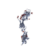 5lfrC  5lfuC 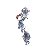 5lfvC  1cs6S  1urlS 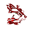 2yd6S 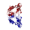 3p3yS  4frwS S: Starting model for refinement C: citing same article ( |
|---|---|
| Similar structure data |


-Links
 PDBj
PDBj


- Assembly
| Deposited unit | 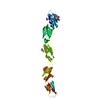
| ||||||||
|---|---|---|---|---|---|---|---|---|---|
| 1 | 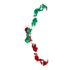
| ||||||||
| Unit cell |
|
-Components
| #1: Protein | Mass: 55192.984 Da / Num. of mol.: 1 Source method: isolated from a genetically manipulated source Details: The two C-terminal residues of the construct and the cloning sites and purification tag are not observed in the electron density, probably because of disorder Source: (gene. exp.)  Homo sapiens (human) / Variant (production host): GnTI-/- and EBNA1-expressing / References: UniProt: P20917 Homo sapiens (human) / Variant (production host): GnTI-/- and EBNA1-expressing / References: UniProt: P20917 | ||
|---|---|---|---|
| #2: Polysaccharide | alpha-L-fucopyranose-(1-6)-2-acetamido-2-deoxy-beta-D-glucopyranose Source method: isolated from a genetically manipulated source | ||
| #3: Polysaccharide | N-acetyl-alpha-neuraminic acid-(2-3)-beta-D-galactopyranose-(1-3)-2-acetamido-2-deoxy-beta-D-galactopyranose Source method: isolated from a genetically manipulated source | ||
| #4: Sugar | ChemComp-MAN /   Type: D-saccharide, alpha linking / Mass: 180.156 Da / Num. of mol.: 1 Type: D-saccharide, alpha linking / Mass: 180.156 Da / Num. of mol.: 1Source method: isolated from a genetically manipulated source Formula: C6H12O6 | ||
| #5: Sugar | ChemComp-NAG /   Type: D-saccharide, beta linking / Mass: 221.208 Da / Num. of mol.: 4 Type: D-saccharide, beta linking / Mass: 221.208 Da / Num. of mol.: 4Source method: isolated from a genetically manipulated source Formula: C8H15NO6 Has protein modification | Y | |
-Experimental details
-Experiment
| Experiment | Method: X-RAY DIFFRACTION / Number of used crystals: 1 |
|---|
- Sample preparation
| Crystal | Density Matthews: 12.7 Å3/Da / Density % sol: 90.33 % |
|---|---|
| Crystal grow | Temperature: 291 K / Method: vapor diffusion, sitting drop / pH: 7 Details: 100 mM NaCl, 20 mM Tris/HCl pH 7.0, 7.7 % PEG 4000 (w/v) |
-Data collection
| Diffraction | Mean temperature: 100 K |
|---|---|
| Diffraction source | Source: SYNCHROTRON / Site: SLS  / Beamline: X06SA / Wavelength: 0.99998 Å / Beamline: X06SA / Wavelength: 0.99998 Å |
| Detector | Type: DECTRIS PILATUS3 6M / Detector: PIXEL / Date: Aug 22, 2013 |
| Radiation | Protocol: SINGLE WAVELENGTH / Monochromatic (M) / Laue (L): M / Scattering type: x-ray |
| Radiation wavelength | Wavelength: 0.99998 Å / Relative weight: 1 |
| Reflection | Resolution: 3.8→52.7 Å / Num. obs: 27623 / % possible obs: 100 % / Redundancy: 9.6 % / CC1/2: 0.997 / Rmerge(I) obs: 0.233 / Net I/σ(I): 9.3 |
| Reflection shell | Resolution: 3.8→4.03 Å / Redundancy: 9.7 % / Rmerge(I) obs: 1.685 / Mean I/σ(I) obs: 1.6 / CC1/2: 0.565 / % possible all: 100 |
- Processing
| Software |
| ||||||||||||||||||||||||||||||||||||||||||||||||||||||||||||||||||||||||||||||||||||||||||||||||||||||||||||||||||||||||||||||||||||||||||||||||||||||
|---|---|---|---|---|---|---|---|---|---|---|---|---|---|---|---|---|---|---|---|---|---|---|---|---|---|---|---|---|---|---|---|---|---|---|---|---|---|---|---|---|---|---|---|---|---|---|---|---|---|---|---|---|---|---|---|---|---|---|---|---|---|---|---|---|---|---|---|---|---|---|---|---|---|---|---|---|---|---|---|---|---|---|---|---|---|---|---|---|---|---|---|---|---|---|---|---|---|---|---|---|---|---|---|---|---|---|---|---|---|---|---|---|---|---|---|---|---|---|---|---|---|---|---|---|---|---|---|---|---|---|---|---|---|---|---|---|---|---|---|---|---|---|---|---|---|---|---|---|---|---|---|
| Refinement | Method to determine structure: MOLECULAR REPLACEMENT Starting model: 1URL, 4FRW, 1CS6, 3P3Y, 2YD6 Resolution: 3.8→52.701 Å / SU ML: 0.51 / Cross valid method: FREE R-VALUE / σ(F): 1.34 / Phase error: 25.3 / Stereochemistry target values: ML
| ||||||||||||||||||||||||||||||||||||||||||||||||||||||||||||||||||||||||||||||||||||||||||||||||||||||||||||||||||||||||||||||||||||||||||||||||||||||
| Solvent computation | Shrinkage radii: 0.9 Å / VDW probe radii: 1.11 Å / Solvent model: FLAT BULK SOLVENT MODEL | ||||||||||||||||||||||||||||||||||||||||||||||||||||||||||||||||||||||||||||||||||||||||||||||||||||||||||||||||||||||||||||||||||||||||||||||||||||||
| Refinement step | Cycle: LAST / Resolution: 3.8→52.701 Å
| ||||||||||||||||||||||||||||||||||||||||||||||||||||||||||||||||||||||||||||||||||||||||||||||||||||||||||||||||||||||||||||||||||||||||||||||||||||||
| Refine LS restraints |
| ||||||||||||||||||||||||||||||||||||||||||||||||||||||||||||||||||||||||||||||||||||||||||||||||||||||||||||||||||||||||||||||||||||||||||||||||||||||
| LS refinement shell |
| ||||||||||||||||||||||||||||||||||||||||||||||||||||||||||||||||||||||||||||||||||||||||||||||||||||||||||||||||||||||||||||||||||||||||||||||||||||||
| Refinement TLS params. | Method: refined / Refine-ID: X-RAY DIFFRACTION
| ||||||||||||||||||||||||||||||||||||||||||||||||||||||||||||||||||||||||||||||||||||||||||||||||||||||||||||||||||||||||||||||||||||||||||||||||||||||
| Refinement TLS group |
|
