 Movie
Movie Controller

[English] 日本語
 Yorodumi
- PDB-5n1q: METHYL-COENZYME M REDUCTASE III FROM METHANOTHERMOCOCCUS THERMOLI... -
+ Open data

- Basic information
| Entry | Database: PDB / ID: 5n1q | ||||||
|---|---|---|---|---|---|---|---|
| Title | METHYL-COENZYME M REDUCTASE III FROM METHANOTHERMOCOCCUS THERMOLITHOTROPHICUS AT 1.9 A RESOLUTION | ||||||
 Components Components | (METHYL-COENZYME M REDUCTASE III FROM METHANOTHERMOCOCCUS THERMOLITHOTROPHICUS SUBUNIT ...) x 3 | ||||||
 Keywords Keywords | TRANSFERASE / POST-TRANSLATIONAL MODIFICATION / BINDING SITES / CATALYSIS / COENZYMES / DISULFIDES / HYDROGEN / HYDROGEN BONDING / LIGANDS / MESNA / METALLOPORPHYRINS / METHANE / METHANOCOCCALES / NICKEL / OXIDATION-REDUCTION / OXIDOREDUCTASES / PHOSPHOTHREONINE / PROTEIN CONFORMATION / PROTEIN FOLDING / PROTEIN STRUCTURE / THERMOPHILE / AUTOTROPH | ||||||
| Function / homology |  Function and homology information Function and homology informationcoenzyme-B sulfoethylthiotransferase / coenzyme-B sulfoethylthiotransferase activity / methanogenesis / metal ion binding / cytoplasm Similarity search - Function | ||||||
| Biological species |  Methanothermococcus thermolithotrophicus DSM 2095 (archaea) Methanothermococcus thermolithotrophicus DSM 2095 (archaea) | ||||||
| Method |  X-RAY DIFFRACTION / SYNCHROTRON / MOLECULAR REPLACEMENT / Resolution: 1.9 Å X-RAY DIFFRACTION / SYNCHROTRON / MOLECULAR REPLACEMENT / Resolution: 1.9 Å | ||||||
 Authors Authors | Wagner, T. / Wegner, C.E. / Ermler, U. / Shima, S. | ||||||
 Citation Citation | Journal: J.Bacteriol. / Year: 2017 Title: Phylogenetic and Structural Comparisons of the Three Types of Methyl Coenzyme M Reductase from Methanococcales and Methanobacteriales. Authors: Wagner, T. / Wegner, C.E. / Kahnt, J. / Ermler, U. / Shima, S. | ||||||
| History |
|
- Structure visualization
| Structure viewer | Molecule: MolmilJmol/JSmol |
|---|
- Downloads & links
-Download
| PDBx/mmCIF format | 5n1q.cif.gz | 988.3 KB | Display | PDBx/mmCIF format |
|---|---|---|---|---|
| PDB format | pdb5n1q.ent.gz | 815.9 KB | Display | PDB format |
| PDBx/mmJSON format | 5n1q.json.gz | Tree view | PDBx/mmJSON format | |
| Others |  Other downloads Other downloads |
-Validation report
| Arichive directory | https://data.pdbj.org/pub/pdb/validation_reports/n1/5n1qftp://data.pdbj.org/pub/pdb/validation_reports/n1/5n1q | HTTPS FTP |
|---|
-Related structure data

-Links
 PDBj
PDBj
- Assembly
| Deposited unit | 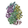
| ||||||||
|---|---|---|---|---|---|---|---|---|---|
| 1 |
| ||||||||
| Unit cell |
|
-Components
-METHYL-COENZYME M REDUCTASE III FROM METHANOTHERMOCOCCUS THERMOLITHOTROPHICUS SUBUNIT ... , 3 types, 6 molecules ADBECF
| #1: Protein | Mass: 61194.961 Da / Num. of mol.: 2 / Source method: isolated from a natural source Details: IN CHAIN A, D RESIDUE 261 IS A N1-METHYLHISTIDINE. RESIDUE 275 IS A C5-(S)-METHYLARGININE. RESIDUE 403 IS A C2-(S)-METHYLGLUTAMINE. RESIDUE 448 IS A THIOGLYCINE. Source: (natural) Methanothermococcus thermolithotrophicus DSM 2095 (archaea)Plasmid details: DSMZ References: UniProt: A0A247D6X3*PLUS, coenzyme-B sulfoethylthiotransferase #2: Protein | Mass: 47362.211 Da / Num. of mol.: 2 / Source method: isolated from a natural source Source: (natural) Methanothermococcus thermolithotrophicus DSM 2095 (archaea)Plasmid details: DSMZ References: UniProt: A0A247D6X4*PLUS, coenzyme-B sulfoethylthiotransferase #3: Protein | Mass: 30368.537 Da / Num. of mol.: 2 / Source method: isolated from a natural source Source: (natural) Methanothermococcus thermolithotrophicus DSM 2095 (archaea)Plasmid details: DSMZ References: UniProt: A0A247D6X5*PLUS, coenzyme-B sulfoethylthiotransferase |
|---|
-Non-polymers , 7 types, 1294 molecules 


| #4: Chemical |  Mass: 343.334 Da / Num. of mol.: 2 / Source method: obtained synthetically / Formula: C11H22NO7PS Mass: 343.334 Da / Num. of mol.: 2 / Source method: obtained synthetically / Formula: C11H22NO7PS#5: Chemical |  Mass: 906.580 Da / Num. of mol.: 2 / Source method: obtained synthetically / Formula: C42H51N6NiO13 Mass: 906.580 Da / Num. of mol.: 2 / Source method: obtained synthetically / Formula: C42H51N6NiO13#6: Chemical | ChemComp-K / |  Mass: 39.098 Da / Num. of mol.: 1 / Source method: obtained synthetically / Formula: K Mass: 39.098 Da / Num. of mol.: 1 / Source method: obtained synthetically / Formula: K#7: Chemical |  Mass: 142.197 Da / Num. of mol.: 2 / Source method: obtained synthetically / Formula: C2H6O3S2 Mass: 142.197 Da / Num. of mol.: 2 / Source method: obtained synthetically / Formula: C2H6O3S2#8: Chemical |  Mass: 92.094 Da / Num. of mol.: 3 / Source method: obtained synthetically / Formula: C3H8O3 Mass: 92.094 Da / Num. of mol.: 3 / Source method: obtained synthetically / Formula: C3H8O3#9: Chemical | ChemComp-MG / |  Mass: 24.305 Da / Num. of mol.: 1 / Source method: obtained synthetically / Formula: Mg Mass: 24.305 Da / Num. of mol.: 1 / Source method: obtained synthetically / Formula: Mg#10: Water | ChemComp-HOH / | Mass: 18.015 Da / Num. of mol.: 1283 / Source method: isolated from a natural source / Formula: H2O |
|---|
-Experimental details
-Experiment
| Experiment | Method: X-RAY DIFFRACTION / Number of used crystals: 1 |
|---|
- Sample preparation
| Crystal | Density Matthews: 2.19 Å3/Da / Density % sol: 43.8 % / Description: Yellow brick |
|---|---|
| Crystal grow | Temperature: 291.15 K / Method: vapor diffusion, sitting drop / pH: 7.6 Details: 1 ul of MCR III from M. thermolithotrophicus with a concentration of 35 mg/ml was mixed with 1 ul of reservoir solution. Best crystals with a yellow brick morphology appeared after a few ...Details: 1 ul of MCR III from M. thermolithotrophicus with a concentration of 35 mg/ml was mixed with 1 ul of reservoir solution. Best crystals with a yellow brick morphology appeared after a few days in 19% (w/v) polyethylene glycol 3350 and 200 mM MgCl2 in the absence of buffer. The crystals were immersed in a solution containing 19% (w/v) PEG 3350 and 200 mM MgCl2, 30% glycerol (v/v) prior to freezing in liquid nitrogen. Temp details: grow between 290 - 293 Kelvin |
-Data collection
| Diffraction | Mean temperature: 100 K |
|---|---|
| Diffraction source | Source: SYNCHROTRON / Site: SLS  / Beamline: X10SA / Wavelength: 0.99979 Å / Beamline: X10SA / Wavelength: 0.99979 Å |
| Detector | Type: DECTRIS PILATUS 6M / Detector: PIXEL / Date: Nov 30, 2015 |
| Radiation | Protocol: SINGLE WAVELENGTH / Monochromatic (M) / Laue (L): M / Scattering type: x-ray |
| Radiation wavelength | Wavelength: 0.99979 Å / Relative weight: 1 |
| Reflection | Resolution: 1.9→46.36 Å / Num. obs: 185011 / % possible obs: 99.2 % / Redundancy: 3.7 % / Biso Wilson estimate: 31.92 Å2 / CC1/2: 0.994 / Rmerge(I) obs: 0.087 / Rpim(I) all: 0.053 / Net I/σ(I): 8.1 |
| Reflection shell | Resolution: 1.9→2 Å / Redundancy: 3.7 % / Rmerge(I) obs: 0.577 / Mean I/σ(I) obs: 1.9 / Num. unique obs: 26774 / CC1/2: 0.804 / Rpim(I) all: 0.346 / % possible all: 98.7 |
- Processing
| Software |
| |||||||||||||||||||||||||||||||||||||||||||||||||||||||||||||||||||||||||||||||||||||||||||||||||||||||||||||||||||||||||||||||||||||||||||||||||||||||||||||||||||||||||||||||
|---|---|---|---|---|---|---|---|---|---|---|---|---|---|---|---|---|---|---|---|---|---|---|---|---|---|---|---|---|---|---|---|---|---|---|---|---|---|---|---|---|---|---|---|---|---|---|---|---|---|---|---|---|---|---|---|---|---|---|---|---|---|---|---|---|---|---|---|---|---|---|---|---|---|---|---|---|---|---|---|---|---|---|---|---|---|---|---|---|---|---|---|---|---|---|---|---|---|---|---|---|---|---|---|---|---|---|---|---|---|---|---|---|---|---|---|---|---|---|---|---|---|---|---|---|---|---|---|---|---|---|---|---|---|---|---|---|---|---|---|---|---|---|---|---|---|---|---|---|---|---|---|---|---|---|---|---|---|---|---|---|---|---|---|---|---|---|---|---|---|---|---|---|---|---|---|---|
| Refinement | Method to determine structure: MOLECULAR REPLACEMENT Starting model: MCR III from Methanotorris formicicus Resolution: 1.9→45.63 Å / Cor.coef. Fo:Fc: 0.9567 / Cor.coef. Fo:Fc free: 0.9485 / SU R Cruickshank DPI: 0.146 / Cross valid method: THROUGHOUT / σ(F): 0 / SU R Blow DPI: 0.147 / SU Rfree Blow DPI: 0.121 / SU Rfree Cruickshank DPI: 0.122
| |||||||||||||||||||||||||||||||||||||||||||||||||||||||||||||||||||||||||||||||||||||||||||||||||||||||||||||||||||||||||||||||||||||||||||||||||||||||||||||||||||||||||||||||
| Displacement parameters | Biso mean: 35.43 Å2
| |||||||||||||||||||||||||||||||||||||||||||||||||||||||||||||||||||||||||||||||||||||||||||||||||||||||||||||||||||||||||||||||||||||||||||||||||||||||||||||||||||||||||||||||
| Refine analyze | Luzzati coordinate error obs: 0.229 Å | |||||||||||||||||||||||||||||||||||||||||||||||||||||||||||||||||||||||||||||||||||||||||||||||||||||||||||||||||||||||||||||||||||||||||||||||||||||||||||||||||||||||||||||||
| Refinement step | Cycle: 1 / Resolution: 1.9→45.63 Å
| |||||||||||||||||||||||||||||||||||||||||||||||||||||||||||||||||||||||||||||||||||||||||||||||||||||||||||||||||||||||||||||||||||||||||||||||||||||||||||||||||||||||||||||||
| Refine LS restraints |
| |||||||||||||||||||||||||||||||||||||||||||||||||||||||||||||||||||||||||||||||||||||||||||||||||||||||||||||||||||||||||||||||||||||||||||||||||||||||||||||||||||||||||||||||
| LS refinement shell | Resolution: 1.9→1.95 Å / Total num. of bins used: 20
| |||||||||||||||||||||||||||||||||||||||||||||||||||||||||||||||||||||||||||||||||||||||||||||||||||||||||||||||||||||||||||||||||||||||||||||||||||||||||||||||||||||||||||||||
| Refinement TLS params. | Method: refined / Refine-ID: X-RAY DIFFRACTION
| |||||||||||||||||||||||||||||||||||||||||||||||||||||||||||||||||||||||||||||||||||||||||||||||||||||||||||||||||||||||||||||||||||||||||||||||||||||||||||||||||||||||||||||||
| Refinement TLS group |
|
