 Movie
Movie Controller

[English] 日本語
 Yorodumi
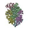
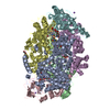
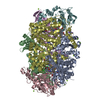
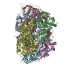
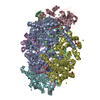
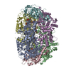
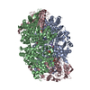
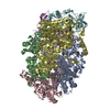
- PDB-3m30: Structural Insight into Methyl-Coenzyme M Reductase Chemistry usi... -
+ Open data

- Basic information
| Entry | Database: PDB / ID: 3m30 | ||||||
|---|---|---|---|---|---|---|---|
| Title | Structural Insight into Methyl-Coenzyme M Reductase Chemistry using Coenzyme B Analogues | ||||||
 Components Components | (Methyl-coenzyme M reductase I subunit ...) x 3 | ||||||
 Keywords Keywords | TRANSFERASE / Methyl-Coenzyme M Reductase / Nickel / Metal-binding / Methanogenesis / Methylation | ||||||
| Function / homology |  Function and homology information Function and homology informationcoenzyme-B sulfoethylthiotransferase / coenzyme-B sulfoethylthiotransferase activity / methanogenesis / metal ion binding / cytoplasm Similarity search - Function | ||||||
| Biological species |   Methanothermobacter marburgensis (archaea) Methanothermobacter marburgensis (archaea) | ||||||
| Method |  X-RAY DIFFRACTION / SYNCHROTRON / FOURIER SYNTHESIS / Resolution: 1.45 Å X-RAY DIFFRACTION / SYNCHROTRON / FOURIER SYNTHESIS / Resolution: 1.45 Å | ||||||
 Authors Authors | Cedervall, P.E. / Dey, M. / Ragsdale, S.W. / Wilmot, C.M. | ||||||
 Citation Citation | Journal: Biochemistry / Year: 2010 Title: Structural insight into methyl-coenzyme M reductase chemistry using coenzyme B analogues. Authors: Cedervall, P.E. / Dey, M. / Pearson, A.R. / Ragsdale, S.W. / Wilmot, C.M. | ||||||
| History |
|
- Structure visualization
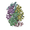
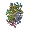
| Structure viewer | Molecule: MolmilJmol/JSmol |
|---|
- Downloads & links
-Download
| PDBx/mmCIF format | 3m30.cif.gz | 574.4 KB | Display | PDBx/mmCIF format |
|---|---|---|---|---|
| PDB format | pdb3m30.ent.gz | 460.6 KB | Display | PDB format |
| PDBx/mmJSON format | 3m30.json.gz | Tree view | PDBx/mmJSON format | |
| Others |  Other downloads Other downloads |
-Validation report
| Arichive directory | https://data.pdbj.org/pub/pdb/validation_reports/m3/3m30ftp://data.pdbj.org/pub/pdb/validation_reports/m3/3m30 | HTTPS FTP |
|---|
-Related structure data


-Links
 PDBj
PDBj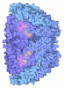
- Assembly
| Deposited unit | 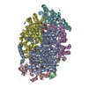
| ||||||||
|---|---|---|---|---|---|---|---|---|---|
| 1 |
| ||||||||
| Unit cell |
|
-Components
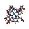
-Methyl-coenzyme M reductase I subunit ... , 3 types, 6 molecules ADBECF
| #1: Protein | Mass: 60508.469 Da / Num. of mol.: 2 / Source method: isolated from a natural source Source: (natural) Methanothermobacter marburgensis (archaea)Strain: Marburg / DSM 2133 References: UniProt: P11558, coenzyme-B sulfoethylthiotransferase #2: Protein | Mass: 47148.477 Da / Num. of mol.: 2 / Source method: isolated from a natural source Source: (natural) Methanothermobacter marburgensis (archaea)Strain: Marburg / DSM 2133 References: UniProt: P11560, coenzyme-B sulfoethylthiotransferase #3: Protein | Mass: 28666.039 Da / Num. of mol.: 2 / Source method: isolated from a natural source Source: (natural) Methanothermobacter marburgensis (archaea)Strain: Marburg / DSM 2133 References: UniProt: P11562, coenzyme-B sulfoethylthiotransferase |
|---|
-Non-polymers , 10 types, 2395 molecules 
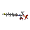
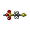
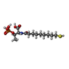


| #4: Chemical | ChemComp-MG /  Mass: 24.305 Da / Num. of mol.: 9 / Source method: obtained synthetically / Formula: Mg Mass: 24.305 Da / Num. of mol.: 9 / Source method: obtained synthetically / Formula: Mg#5: Chemical |  Mass: 906.580 Da / Num. of mol.: 2 / Source method: obtained synthetically / Formula: C42H51N6NiO13 Mass: 906.580 Da / Num. of mol.: 2 / Source method: obtained synthetically / Formula: C42H51N6NiO13#6: Chemical |  Mass: 343.334 Da / Num. of mol.: 2 / Source method: obtained synthetically / Formula: C11H22NO7PS Mass: 343.334 Da / Num. of mol.: 2 / Source method: obtained synthetically / Formula: C11H22NO7PS#7: Chemical |  Mass: 142.197 Da / Num. of mol.: 2 / Source method: obtained synthetically / Formula: C2H6O3S2 Mass: 142.197 Da / Num. of mol.: 2 / Source method: obtained synthetically / Formula: C2H6O3S2#8: Chemical |  Mass: 371.387 Da / Num. of mol.: 2 / Source method: obtained synthetically / Formula: C13H26NO7PS Mass: 371.387 Da / Num. of mol.: 2 / Source method: obtained synthetically / Formula: C13H26NO7PS#9: Chemical |  Mass: 59.044 Da / Num. of mol.: 2 / Source method: obtained synthetically / Formula: C2H3O2 Mass: 59.044 Da / Num. of mol.: 2 / Source method: obtained synthetically / Formula: C2H3O2#10: Chemical | ChemComp-EDO /  Mass: 62.068 Da / Num. of mol.: 6 / Source method: obtained synthetically / Formula: C2H6O2 Mass: 62.068 Da / Num. of mol.: 6 / Source method: obtained synthetically / Formula: C2H6O2#11: Chemical | ChemComp-ZN / |  Mass: 65.409 Da / Num. of mol.: 1 / Source method: obtained synthetically / Formula: Zn Mass: 65.409 Da / Num. of mol.: 1 / Source method: obtained synthetically / Formula: Zn#12: Chemical | ChemComp-PEG / |  Mass: 106.120 Da / Num. of mol.: 1 / Source method: obtained synthetically / Formula: C4H10O3 Mass: 106.120 Da / Num. of mol.: 1 / Source method: obtained synthetically / Formula: C4H10O3#13: Water | ChemComp-HOH / | Mass: 18.015 Da / Num. of mol.: 2368 / Source method: isolated from a natural source / Formula: H2O |
|---|
-Details
| Has protein modification | Y |
|---|
-Experimental details
-Experiment
| Experiment | Method: X-RAY DIFFRACTION / Number of used crystals: 1 |
|---|
- Sample preparation
| Crystal | Density Matthews: 2.18 Å3/Da / Density % sol: 43.47 % |
|---|---|
| Crystal grow | Temperature: 282 K / Method: vapor diffusion, sitting drop Details: PEG 400, magnesium acetate, vapor diffusion, sitting drop, temperature 282K |
-Data collection
| Diffraction | Mean temperature: 100 K |
|---|---|
| Diffraction source | Source: SYNCHROTRON / Site: APS  / Beamline: 14-BM-C / Wavelength: 0.9002 Å / Beamline: 14-BM-C / Wavelength: 0.9002 Å |
| Detector | Type: ADSC QUANTUM 315 / Detector: CCD / Date: Dec 8, 2007 |
| Radiation | Monochromator: Ge(111) / Protocol: SINGLE WAVELENGTH / Monochromatic (M) / Laue (L): M / Scattering type: x-ray |
| Radiation wavelength | Wavelength: 0.9002 Å / Relative weight: 1 |
| Reflection | Resolution: 1.45→50 Å / Num. all: 409587 / Num. obs: 401701 / % possible obs: 98.1 % / Observed criterion σ(F): 1 / Observed criterion σ(I): 3 / Redundancy: 3.5 % / Rmerge(I) obs: 0.056 / Net I/σ(I): 24.283 |
| Reflection shell | Resolution: 1.45→1.5 Å / Redundancy: 3.3 % / Rmerge(I) obs: 0.425 / Mean I/σ(I) obs: 3.167 / % possible all: 95.4 |
- Processing
| Software |
| ||||||||||||||||||||||||||||||||||||||||||||||||||||||||||||||||||||||||||||||||||||||||||||||||||||||||||||||||||||||||||||||||||||||||||||||||||||||||||||||||||||||||||
|---|---|---|---|---|---|---|---|---|---|---|---|---|---|---|---|---|---|---|---|---|---|---|---|---|---|---|---|---|---|---|---|---|---|---|---|---|---|---|---|---|---|---|---|---|---|---|---|---|---|---|---|---|---|---|---|---|---|---|---|---|---|---|---|---|---|---|---|---|---|---|---|---|---|---|---|---|---|---|---|---|---|---|---|---|---|---|---|---|---|---|---|---|---|---|---|---|---|---|---|---|---|---|---|---|---|---|---|---|---|---|---|---|---|---|---|---|---|---|---|---|---|---|---|---|---|---|---|---|---|---|---|---|---|---|---|---|---|---|---|---|---|---|---|---|---|---|---|---|---|---|---|---|---|---|---|---|---|---|---|---|---|---|---|---|---|---|---|---|---|---|---|
| Refinement | Method to determine structure: FOURIER SYNTHESIS / Resolution: 1.45→20.07 Å / Cor.coef. Fo:Fc: 0.977 / Cor.coef. Fo:Fc free: 0.968 / Occupancy max: 1 / Occupancy min: 0 / SU B: 0.948 / SU ML: 0.036 / SU R Cruickshank DPI: 0.056 / Cross valid method: THROUGHOUT / ESU R: 0.057 / ESU R Free: 0.06 / Stereochemistry target values: MAXIMUM LIKELIHOOD / Details: HYDROGENS HAVE BEEN ADDED IN THE RIDING POSITIONS
| ||||||||||||||||||||||||||||||||||||||||||||||||||||||||||||||||||||||||||||||||||||||||||||||||||||||||||||||||||||||||||||||||||||||||||||||||||||||||||||||||||||||||||
| Solvent computation | Ion probe radii: 0.8 Å / Shrinkage radii: 0.8 Å / VDW probe radii: 1.4 Å / Solvent model: MASK | ||||||||||||||||||||||||||||||||||||||||||||||||||||||||||||||||||||||||||||||||||||||||||||||||||||||||||||||||||||||||||||||||||||||||||||||||||||||||||||||||||||||||||
| Displacement parameters | Biso mean: 13.931 Å2
| ||||||||||||||||||||||||||||||||||||||||||||||||||||||||||||||||||||||||||||||||||||||||||||||||||||||||||||||||||||||||||||||||||||||||||||||||||||||||||||||||||||||||||
| Refinement step | Cycle: LAST / Resolution: 1.45→20.07 Å
| ||||||||||||||||||||||||||||||||||||||||||||||||||||||||||||||||||||||||||||||||||||||||||||||||||||||||||||||||||||||||||||||||||||||||||||||||||||||||||||||||||||||||||
| Refine LS restraints |
| ||||||||||||||||||||||||||||||||||||||||||||||||||||||||||||||||||||||||||||||||||||||||||||||||||||||||||||||||||||||||||||||||||||||||||||||||||||||||||||||||||||||||||
| LS refinement shell | Resolution: 1.45→1.484 Å / Total num. of bins used: 20
|
