 Movie
Movie Controller

[English] 日本語
 Yorodumi
- PDB-5fmm: crystal structure of the mid, cap-binding, mid-link and 627 domai... -
+ Open data

- Basic information
| Entry | Database: PDB / ID: 5fmm | ||||||
|---|---|---|---|---|---|---|---|
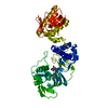
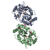
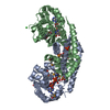
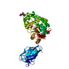
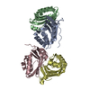
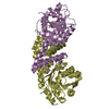
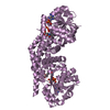
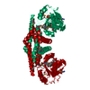
| Title | crystal structure of the mid, cap-binding, mid-link and 627 domains from avian influenza a virus polymerase PB2 subunit bound to M7GTP | ||||||
 Components Components | INFLUENZA A PB2 SUBUNIT | ||||||
 Keywords Keywords | VIRAL PROTEIN / INFLUENZA A PB2-C REGION CONTAINING MID / CAP-BINDING / MID-LINK / 627 AND NLS DOMAINS | ||||||
| Function / homology |  Function and homology information Function and homology informationcap snatching / symbiont-mediated suppression of host mRNA transcription via inhibition of RNA polymerase II activity / 7-methylguanosine mRNA capping / host cell mitochondrion / symbiont-mediated suppression of host cytoplasmic pattern recognition receptor signaling pathway via inhibition of MAVS activity / virion component / symbiont-mediated suppression of host gene expression / viral RNA genome replication / RNA-directed RNA polymerase activity / host cell nucleus ...cap snatching / symbiont-mediated suppression of host mRNA transcription via inhibition of RNA polymerase II activity / 7-methylguanosine mRNA capping / host cell mitochondrion / symbiont-mediated suppression of host cytoplasmic pattern recognition receptor signaling pathway via inhibition of MAVS activity / virion component / symbiont-mediated suppression of host gene expression / viral RNA genome replication / RNA-directed RNA polymerase activity / host cell nucleus / DNA-templated transcription / RNA binding Similarity search - Function | ||||||
| Biological species |   INFLUENZA A VIRUS INFLUENZA A VIRUS | ||||||
| Method |  X-RAY DIFFRACTION / SYNCHROTRON / MOLECULAR REPLACEMENT / Resolution: 2.4 Å X-RAY DIFFRACTION / SYNCHROTRON / MOLECULAR REPLACEMENT / Resolution: 2.4 Å | ||||||
 Authors Authors | Thierry, E. / Pflug, A. / Hart, D. / Cusack, S. | ||||||
 Citation Citation | Journal: Mol.Cell / Year: 2016 Title: Influenza Polymerase Can Adopt an Alternative Configuration Involving a Radical Repacking of Pb2 Domains. Authors: Thierry, E. / Guilligay, D. / Kosinski, J. / Bock, T. / Gaudon, S. / Round, A. / Pflug, A. / Hengrung, N. / El Omari, K. / Baudin, F. / Hart, D.J. / Beck, M. / Cusack, S. | ||||||
| History |
|
- Structure visualization
| Structure viewer | Molecule: MolmilJmol/JSmol |
|---|
- Downloads & links
-Download
| PDBx/mmCIF format | 5fmm.cif.gz | 103.8 KB | Display | PDBx/mmCIF format |
|---|---|---|---|---|
| PDB format | pdb5fmm.ent.gz | 77.9 KB | Display | PDB format |
| PDBx/mmJSON format | 5fmm.json.gz | Tree view | PDBx/mmJSON format | |
| Others |  Other downloads Other downloads |
-Validation report
| Arichive directory | https://data.pdbj.org/pub/pdb/validation_reports/fm/5fmmftp://data.pdbj.org/pub/pdb/validation_reports/fm/5fmm | HTTPS FTP |
|---|
-Related structure data
| Related structure data | 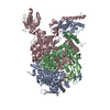 5epiC  5fmlC  5fmqC  5fmzC 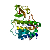 3kc6S  4cb4S  4wsbS C: citing same article ( S: Starting model for refinement |
|---|---|
| Similar structure data |


-Links
 PDBj
PDBj


- Assembly
| Deposited unit | 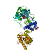
| ||||||||
|---|---|---|---|---|---|---|---|---|---|
| 1 |
| ||||||||
| Unit cell |
|
-Components
| #1: Protein | Mass: 54739.852 Da / Num. of mol.: 1 / Fragment: PB2-C, RESIDUES 247-736 Source method: isolated from a genetically manipulated source Source: (gene. exp.) INFLUENZA A VIRUS / Strain: A/VIETNAM/1203/2004(H5N1) / Production host:  |
|---|---|
| #2: Chemical | ChemComp-MGT /   Mass: 539.223 Da / Num. of mol.: 1 / Source method: obtained synthetically / Formula: C11H20N5O14P3 Mass: 539.223 Da / Num. of mol.: 1 / Source method: obtained synthetically / Formula: C11H20N5O14P3 |
| #3: Chemical | ChemComp-FLC /   Mass: 189.100 Da / Num. of mol.: 1 / Source method: obtained synthetically / Formula: C6H5O7 Mass: 189.100 Da / Num. of mol.: 1 / Source method: obtained synthetically / Formula: C6H5O7 |
| #4: Water | ChemComp-HOH /  Mass: 18.015 Da / Num. of mol.: 125 / Source method: isolated from a natural source / Formula: H2O Mass: 18.015 Da / Num. of mol.: 125 / Source method: isolated from a natural source / Formula: H2O |
| Nonpolymer details | CITRATE ANION (FLC): FROM BUFFER 7N-METHYL-8-HYDROGUANO |
-Experimental details
-Experiment
| Experiment | Method: X-RAY DIFFRACTION / Number of used crystals: 1 |
|---|
- Sample preparation
| Crystal | Density Matthews: 3.1 Å3/Da / Density % sol: 60.2 % / Description: NONE |
|---|---|
| Crystal grow | pH: 5 Details: 0.1 M TRI-SODIUM CITRATE PH 5 10 % POLYETHYLENE GLYCOL MONOMETHYL ETHER 5000 |
-Data collection
| Diffraction | Mean temperature: 100 K |
|---|---|
| Diffraction source | Source: SYNCHROTRON / Site: ESRF  / Beamline: ID29 / Wavelength: 0.97625 / Beamline: ID29 / Wavelength: 0.97625 |
| Detector | Type: DECTRIS PILATUS 2M / Detector: PIXEL / Date: Jan 24, 2015 |
| Radiation | Protocol: SINGLE WAVELENGTH / Monochromatic (M) / Laue (L): M / Scattering type: x-ray |
| Radiation wavelength | Wavelength: 0.97625 Å / Relative weight: 1 |
| Reflection | Resolution: 2.4→50 Å / Num. obs: 26542 / % possible obs: 100 % / Observed criterion σ(I): 0 / Redundancy: 10.5 % / Rmerge(I) obs: 0.15 / Net I/σ(I): 12.2 |
| Reflection shell | Resolution: 2.4→2.48 Å / Redundancy: 10.5 % / Rmerge(I) obs: 1.31 / Mean I/σ(I) obs: 2.04 / % possible all: 100 |
- Processing
| Software |
| ||||||||||||||||||||||||||||||||||||||||||||||||||||||||||||||||||||||||||||||||||||||||||||||||||||||||||||||||||||||||||||||||||||||||||||||||||||||||||||||||||||||||||||||||||||||
|---|---|---|---|---|---|---|---|---|---|---|---|---|---|---|---|---|---|---|---|---|---|---|---|---|---|---|---|---|---|---|---|---|---|---|---|---|---|---|---|---|---|---|---|---|---|---|---|---|---|---|---|---|---|---|---|---|---|---|---|---|---|---|---|---|---|---|---|---|---|---|---|---|---|---|---|---|---|---|---|---|---|---|---|---|---|---|---|---|---|---|---|---|---|---|---|---|---|---|---|---|---|---|---|---|---|---|---|---|---|---|---|---|---|---|---|---|---|---|---|---|---|---|---|---|---|---|---|---|---|---|---|---|---|---|---|---|---|---|---|---|---|---|---|---|---|---|---|---|---|---|---|---|---|---|---|---|---|---|---|---|---|---|---|---|---|---|---|---|---|---|---|---|---|---|---|---|---|---|---|---|---|---|---|
| Refinement | Method to determine structure: MOLECULAR REPLACEMENT Starting model: PDB ENTRIES 4WSB, 4CB4, 3KC6 Resolution: 2.4→84.24 Å / Cor.coef. Fo:Fc: 0.941 / Cor.coef. Fo:Fc free: 0.937 / SU B: 7.261 / SU ML: 0.164 / Cross valid method: THROUGHOUT / ESU R: 0.279 / ESU R Free: 0.209 / Stereochemistry target values: MAXIMUM LIKELIHOOD Details: HYDROGENS HAVE BEEN ADDED IN THE RIDING POSITIONS. U VALUES REFINED INDIVIDUALLY
| ||||||||||||||||||||||||||||||||||||||||||||||||||||||||||||||||||||||||||||||||||||||||||||||||||||||||||||||||||||||||||||||||||||||||||||||||||||||||||||||||||||||||||||||||||||||
| Solvent computation | Ion probe radii: 0.8 Å / Shrinkage radii: 0.8 Å / VDW probe radii: 1.2 Å / Solvent model: MASK | ||||||||||||||||||||||||||||||||||||||||||||||||||||||||||||||||||||||||||||||||||||||||||||||||||||||||||||||||||||||||||||||||||||||||||||||||||||||||||||||||||||||||||||||||||||||
| Displacement parameters | Biso mean: 75.89 Å2
| ||||||||||||||||||||||||||||||||||||||||||||||||||||||||||||||||||||||||||||||||||||||||||||||||||||||||||||||||||||||||||||||||||||||||||||||||||||||||||||||||||||||||||||||||||||||
| Refinement step | Cycle: LAST / Resolution: 2.4→84.24 Å
| ||||||||||||||||||||||||||||||||||||||||||||||||||||||||||||||||||||||||||||||||||||||||||||||||||||||||||||||||||||||||||||||||||||||||||||||||||||||||||||||||||||||||||||||||||||||
| Refine LS restraints |
|